Time-dependent diffusion in undulating structures: Impact on axon diameter estimation
Publication
Metrics
AI Quick Summary
This study investigates the impact of axon undulation on diameter estimation using diffusion MRI, finding that the straight-cylinder model overestimates diameter due to the undulation amplitude and microscopic orientation dispersion. The undulating thin-fiber model reveals differences in high-frequency diffusion spectra compared to straight cylinders, suggesting the need to account for axon trajectory when interpreting dMRI data.
Paper Preview
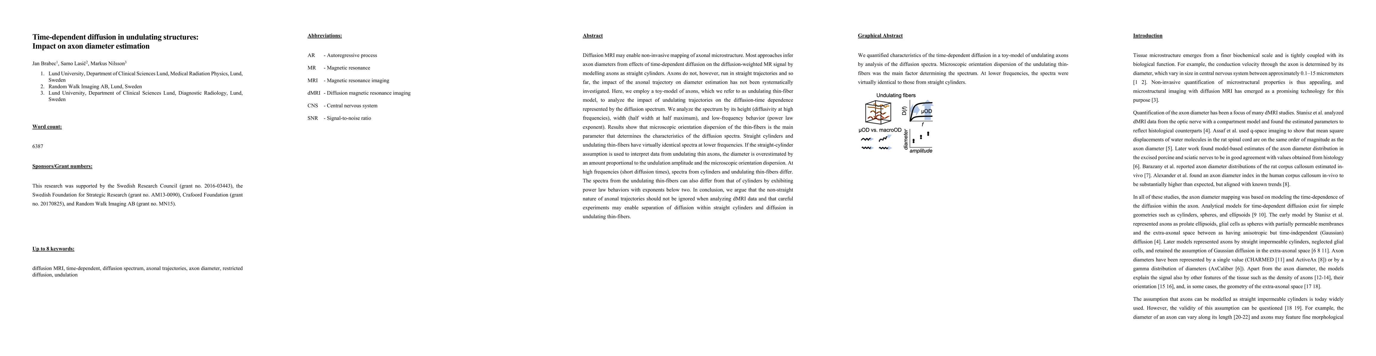
Abstract
Diffusion MRI may enable non-invasive mapping of axonal microstructure. Most approaches infer axon diameters from effects of time-dependent diffusion on the diffusion-weighted MR signal by modelling axons as straight cylinders. Axons do not, however, run in straight trajectories and so far, the impact of the axonal trajectory on diameter estimation has not been systematically investigated. Here, we employ a toy-model of axons, which we refer to as undulating thin-fiber model, to analyze the impact of undulating trajectories on the diffusion-time dependence represented by the diffusion spectrum. We analyze the spectrum by its height (diffusivity at high frequencies), width (half width at half maximum), and low-frequency behavior (power law exponent). Results show that microscopic orientation dispersion of the thin-fibers is the main parameter that determines the characteristics of the diffusion spectra. Straight cylinders and undulating thin-fibers have virtually identical spectra at lower frequencies. If the straight-cylinder assumption is used to interpret data from undulating thin axons, the diameter is overestimated by an amount proportional to the undulation amplitude and the microscopic orientation dispersion. At high frequencies (short diffusion times), spectra from cylinders and undulating thin-fibers differ. The spectra from the undulating thin-fibers can also differ from that of cylinders by exhibiting power law behaviors with exponents below two. In conclusion, we argue that the non-straight nature of axonal trajectories should not be ignored when analyzing dMRI data and that careful experiments may enable separation of diffusion within straight cylinders and diffusion in undulating thin-fibers.
AI Key Findings
Get AI-generated insights about this paper's methodology, results, significance, and more — seven facets brought into focus.
Impact
Paper Details
PDF Preview
Key Terms
Citation Network
Current paper (gray), citations (green), references (blue)
Display is limited for performance on very large graphs.
Discussion 0