Publication
Metrics
AI Quick Summary
This paper introduces a time-multiplexed structured illumination technique using a DMD for optical diffraction tomography, which controls various spatial frequencies to eliminate unwanted diffracted beams, improving image quality. The method was demonstrated to reconstruct 3-D refractive index distributions with high sensitivity and provide biochemical and morphological insights from biological samples.
Paper Preview
Abstract
We present a novel illumination control technique for optical diffraction tomography (ODT). Various spatial frequencies of beam illumination were controlled by displaying time-averaged sinusoidal patterns using a digital micromirror device (DMD). Compared to the previous method using binary Lee holograms, the present method eliminates unwanted diffracted beams which may deteriorate the image quality of the ODT. We demonstrated the capability of the present method by reconstructing three-dimensional refractive index (RI) distributions of various samples, with high RI sensitivity (\sigma_\Delta n = 3.15 +/- 10-4), and reconstructing 3-D RI tomograms of biological samples, which provided quantitative biochemical and morphological information about the samples.
AI Key Findings
Get AI-generated insights about this paper's methodology, results, significance, and more — seven facets brought into focus.
Impact
Paper Details
PDF Preview
Key Terms
Citation Network
Current paper (gray), citations (green), references (blue)
Display is limited for performance on very large graphs.
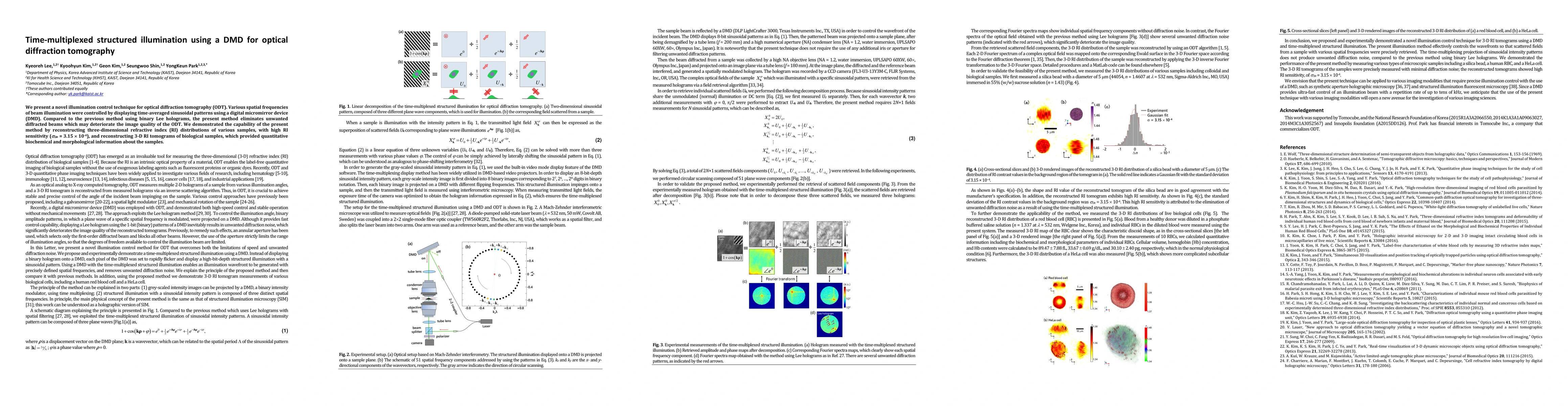
Discussion 0