Summary
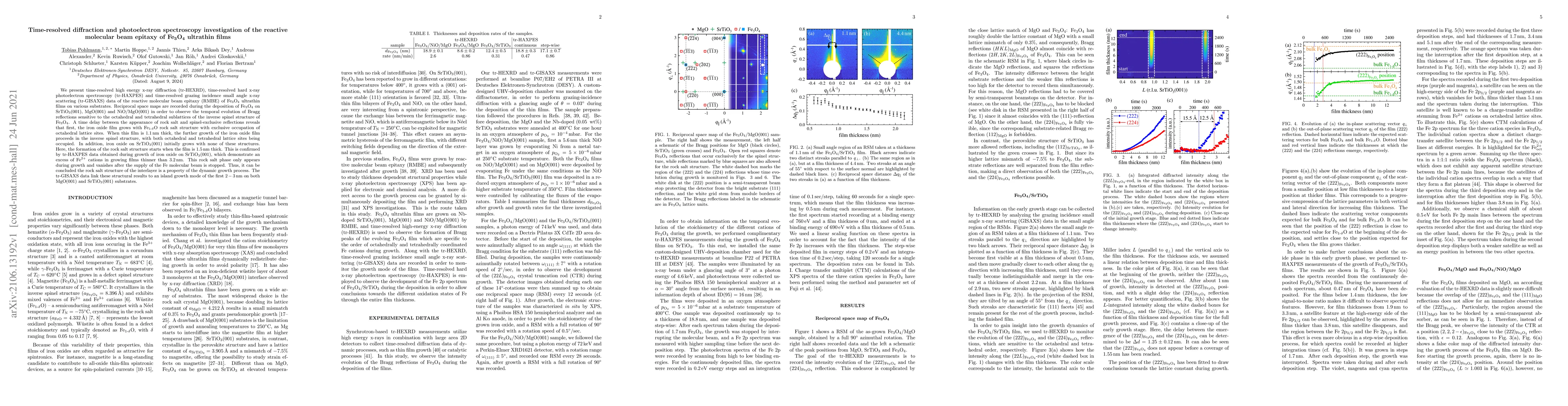
We present time-resolved high energy x-ray diffraction (tr-HEXRD), time-resolved hard x-ray photoelectron spectroscopy (tr-HAXPES) and time-resolved grazing incidence small angle x-ray scattering (tr-GISAXS) data of the reactive molecular beam epitaxy (RMBE) of $\mathrm{Fe_3O_4}$ ultrathin films on various substrates. Reciprocal space maps are recorded during the deposition of $\mathrm{Fe_3O_4}$ on $\mathrm{SrTiO_3(001)}$, MgO(001) and NiO/MgO(001) in order to observe the temporal evolution of Bragg reflections sensitive to the octahedral and tetrahedral sublattices of the inverse spinel structure of $\mathrm{Fe_3O_4}$. A time delay between the appearance of rock salt and spinel-exclusive reflections reveals that first, the iron oxide film grows with $\mathrm{Fe_{1-\delta}O}$ rock salt structure with exclusive occupation of octahedral lattice sites. When this film is 1.1$\,$nm thick, the further growth of the iron oxide film proceeds in the inverse spinel structure, with both octahedral and tetrahedral lattice sites being occupied. In addition, iron oxide on $\mathrm{SrTiO_3(001)}$ initially grows with none of these structures. Here, the formation of the rock salt structure starts when the film is 1.5$\,$nm thick. This is confirmed by tr-HAXPES data obtained during growth of iron oxide on $\mathrm{SrTiO_3(001)}$, which demonstrate an excess of $\mathrm{Fe^{2+}}$ cations in growing films thinner than 3.2$\,$nm. This rock salt phase only appears during growth and vanishes after the supply of the Fe molecular beam is stopped. Thus, it can be concluded the rock salt structure of the interlayer is a property of the dynamic growth process. The tr-GISAXS data link these structural results to an island growth mode of the first 2-3$\,$nm on both MgO(001) and $\mathrm{SrTiO_3(001)}$ substrates.
AI Key Findings
Get AI-generated insights about this paper's methodology, results, and significance.
Paper Details
PDF Preview
Key Terms
Citation Network
Current paper (gray), citations (green), references (blue)
Display is limited for performance on very large graphs.
Similar Papers
Found 4 papersSuperconducting Phase of $\mathrm{Ti_xO_y}$ Thin Films Grown by Molecular Beam Epitaxy
Divine P. Kumah, Xuanyi Zhang, Zhan Zhang et al.
| Title | Authors | Year | Actions |
|---|
Comments (0)