TJDR: A High-Quality Diabetic Retinopathy Pixel-Level Annotation Dataset
Publication
Metrics
AI Quick Summary
This paper introduces TJDR, a high-quality pixel-level annotation dataset for diabetic retinopathy (DR) comprising 561 color fundus images from Tongji Hospital. The dataset aims to enhance DR lesion segmentation research, featuring annotations of four DR lesions by experienced ophthalmologists and released to support further advancements in the field.
Paper Preview
Abstract
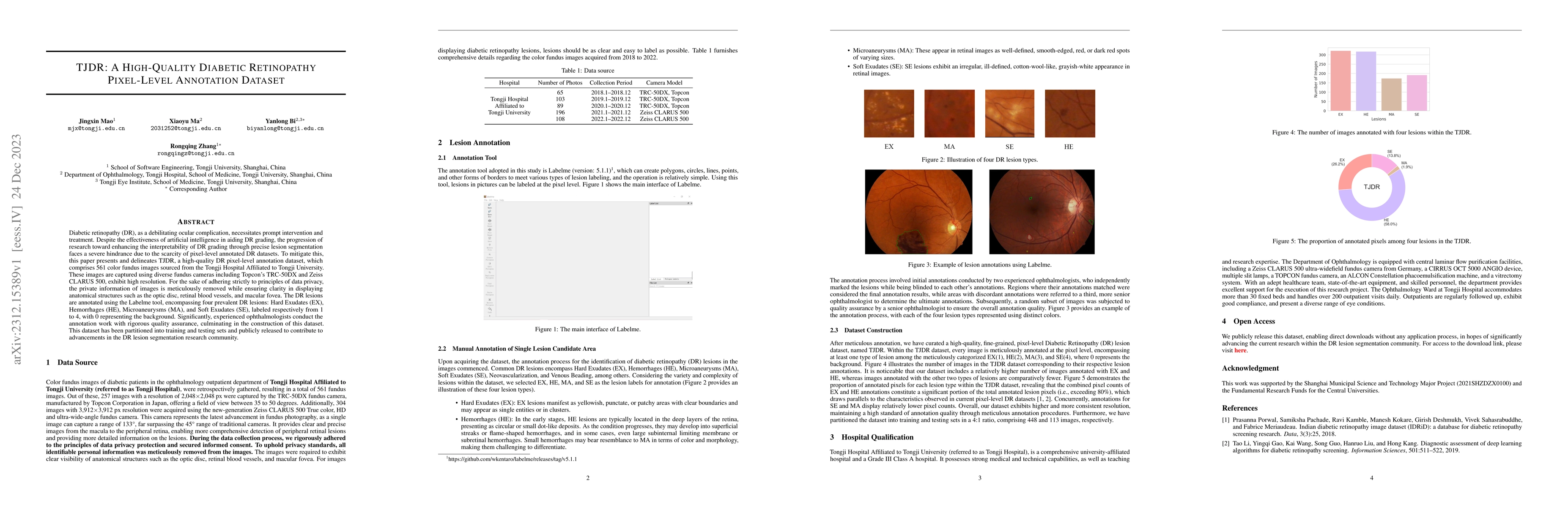
Diabetic retinopathy (DR), as a debilitating ocular complication, necessitates prompt intervention and treatment. Despite the effectiveness of artificial intelligence in aiding DR grading, the progression of research toward enhancing the interpretability of DR grading through precise lesion segmentation faces a severe hindrance due to the scarcity of pixel-level annotated DR datasets. To mitigate this, this paper presents and delineates TJDR, a high-quality DR pixel-level annotation dataset, which comprises 561 color fundus images sourced from the Tongji Hospital Affiliated to Tongji University. These images are captured using diverse fundus cameras including Topcon's TRC-50DX and Zeiss CLARUS 500, exhibit high resolution. For the sake of adhering strictly to principles of data privacy, the private information of images is meticulously removed while ensuring clarity in displaying anatomical structures such as the optic disc, retinal blood vessels, and macular fovea. The DR lesions are annotated using the Labelme tool, encompassing four prevalent DR lesions: Hard Exudates (EX), Hemorrhages (HE), Microaneurysms (MA), and Soft Exudates (SE), labeled respectively from 1 to 4, with 0 representing the background. Significantly, experienced ophthalmologists conduct the annotation work with rigorous quality assurance, culminating in the construction of this dataset. This dataset has been partitioned into training and testing sets and publicly released to contribute to advancements in the DR lesion segmentation research community.
AI Key Findings
Get AI-generated insights about this paper's methodology, results, significance, and more — seven facets brought into focus.
Impact
Paper Details
Authors
PDF Preview
Key Terms
Citation Network
Current paper (gray), citations (green), references (blue)
Display is limited for performance on very large graphs.
Discussion 0