Towards Automatic Embryo Staging in 3D+T Microscopy Images using Convolutional Neural Networks and PointNets
Publication
Metrics
AI Quick Summary
This paper explores automatic staging of embryonic development using convolutional neural networks and PointNets on 3D+T microscopy images. Both methods show promise with average deviations of 21-34 minutes on real zebrafish embryos, and less than 7 minutes on simulated data.
Paper Preview
Abstract
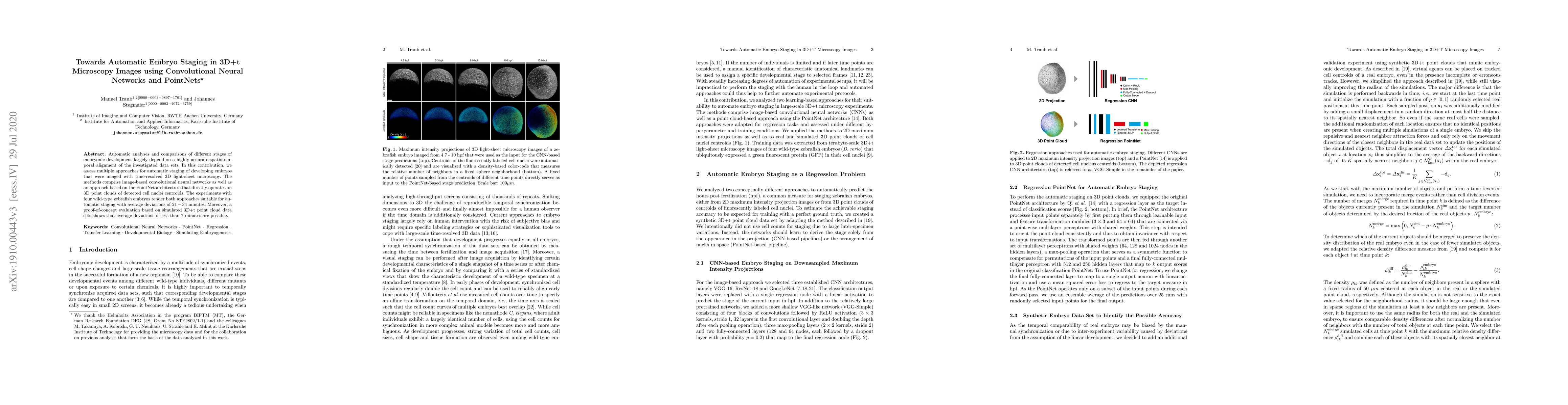
Automatic analyses and comparisons of different stages of embryonic development largely depend on a highly accurate spatiotemporal alignment of the investigated data sets. In this contribution, we assess multiple approaches for automatic staging of developing embryos that were imaged with time-resolved 3D light-sheet microscopy. The methods comprise image-based convolutional neural networks as well as an approach based on the PointNet architecture that directly operates on 3D point clouds of detected cell nuclei centroids. The experiments with four wild-type zebrafish embryos render both approaches suitable for automatic staging with average deviations of 21 - 34 minutes. Moreover, a proof-of-concept evaluation based on simulated 3D+t point cloud data sets shows that average deviations of less than 7 minutes are possible.
AI Key Findings
Get AI-generated insights about this paper's methodology, results, significance, and more — seven facets brought into focus.
Impact
Paper Details
PDF Preview
Key Terms
Citation Network
Current paper (gray), citations (green), references (blue)
Display is limited for performance on very large graphs.
Discussion 0