Authors
Publication
Metrics
Quick Actions
AI Quick Summary
This paper develops automated workflows for structural discovery in STEM, employing active learning and Bayesian optimization to identify regions of interest. It explores the balance between a priori knowledge and experimental data, showcasing methods for image data descriptors and deep learning segmentation, and applies these workflows to various material features.
Quick Answers
What is "Towards Automating Structural Discovery in Scanning Transmission Electron Microscopy" about?
This paper develops automated workflows for structural discovery in STEM, employing active learning and Bayesian optimization to identify regions of interest. It explores the balance between a priori knowledge and experimental data, showcasing methods for image data descriptors and deep learning segmentation, and applies these workflows to various material features.
What methodology did the authors use?
The research explores automated experiment workflows for Scanning Transmission Electron Microscopy (STEM) using a combination of deep convolutional neural networks (DCNN) for semantic segmentation and variational autoencoders (VAE) for encoding. Bayesian optimization (BO) is employed for rapid exploration of sample space for features of interest, with latent variables or components selected during training and automated experimental stages. More in Methodology →
What are the key results?
Workflow 1, using sliding window FFT and lower-level segmentation, proved robust for well-defined features of interest. — Workflow 3, combining DCNN and VAE, performed well for atomic-scale features. More in Key Results →
Why is this work significant?
This research is significant as it lays the groundwork for automated experiments in STEM and Scanning Probe Microscopy (SPM), addressing the need for machine learning algorithms to guide the selection of objects of interest, considering the specificity and generic features of the systems. More in Significance →
What are the main limitations?
The performance of a pre-trained DCNN may not work if the collected image is not within the distribution of data used for training. — Data manipulation for BO during microscope operation can be challenging and time-consuming without a field-programmable gate array (FPGA) connected directly to the microscope. More in Limitations →
Abstract
Scanning transmission electron microscopy (STEM) is now the primary tool for exploring functional materials on the atomic level. Often, features of interest are highly localized in specific regions in the material, such as ferroelectric domain walls, extended defects, or second phase inclusions. Selecting regions to image for structural and chemical discovery via atomically resolved imaging has traditionally proceeded via human operators making semi-informed judgements on sampling locations and parameters. Recent efforts at automation for structural and physical discovery have pointed towards the use of "active learning" methods that utilize Bayesian optimization with surrogate models to quickly find relevant regions of interest. Yet despite the potential importance of this direction, there is a general lack of certainty in selecting relevant control algorithms and how to balance a priori knowledge of the material system with knowledge derived during experimentation. Here we address this gap by developing the automated experiment workflows with several combinations to both illustrate the effects of these choices and demonstrate the tradeoffs associated with each in terms of accuracy, robustness, and susceptibility to hyperparameters for structural discovery. We discuss possible methods to build descriptors using the raw image data and deep learning based semantic segmentation, as well as the implementation of variational autoencoder based 
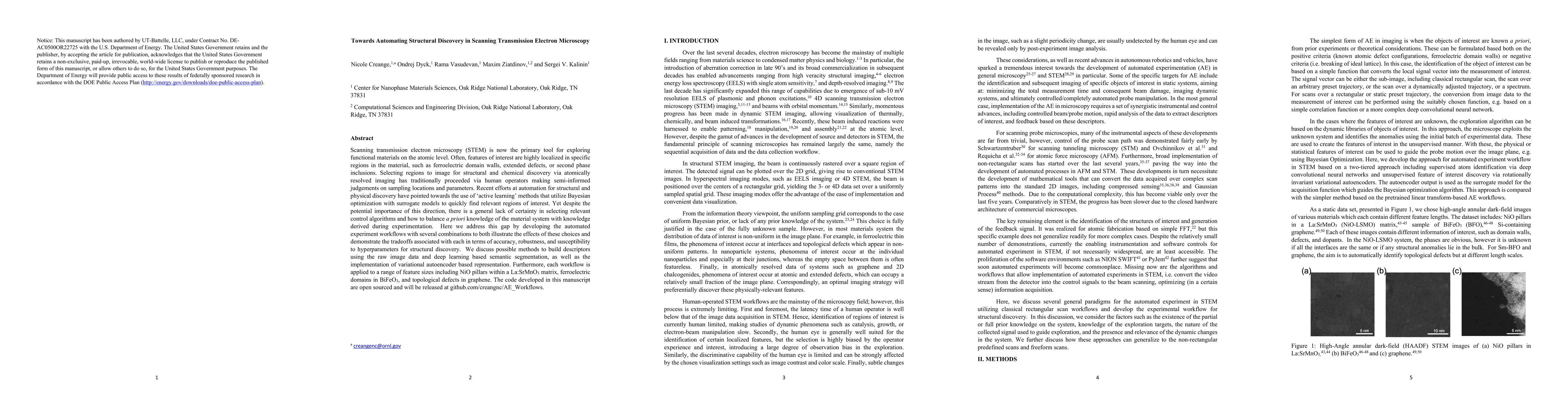
representation. Furthermore, each workflow is applied to a range of feature sizes including NiO pillars within a La:SrMnO$_3$ matrix, ferroelectric domains in BiFeO$_3$, and topological defects in graphene. The code developed in this manuscript are open sourced and will be released at github.com/creangnc/AE_Workflows.
AI Key Findings
Generated Sep 02, 2025
Methodology — What approach did the authors take?
The research explores automated experiment workflows for Scanning Transmission Electron Microscopy (STEM) using a combination of deep convolutional neural networks (DCNN) for semantic segmentation and variational autoencoders (VAE) for encoding. Bayesian optimization (BO) is employed for rapid exploration of sample space for features of interest, with latent variables or components selected during training and automated experimental stages.
Key Results — What are the main findings?
- Workflow 1, using sliding window FFT and lower-level segmentation, proved robust for well-defined features of interest.
- Workflow 3, combining DCNN and VAE, performed well for atomic-scale features.
- The use of BO allowed for efficient data collection, with most points collected in regions of interest, reducing beam damage and focusing on features of interest.
- The workflows demonstrated varying degrees of effectiveness depending on feature length and dataset.
- The study suggests that parameter tuning and main network training should be performed a-priori for time-efficiency, while fine-tuning of the DCNN network could be done within the workflow.
Significance — Why does this research matter?
This research is significant as it lays the groundwork for automated experiments in STEM and Scanning Probe Microscopy (SPM), addressing the need for machine learning algorithms to guide the selection of objects of interest, considering the specificity and generic features of the systems.
Technical Contribution — What is the technical contribution?
The paper presents automated experiment workflows for STEM that integrate DCNN semantic segmentation, VAE encoding, and BO, providing a gateway for rapid feature discovery, minimized beam damage, and data collection primarily focused on features of interest.
Novelty — What is new about this work?
This work stands out by combining DCNN, VAE, and BO for automated feature discovery in STEM, offering a flexible and robust framework that can be adapted to various systems and feature sizes, while also addressing the need for reduced beam damage and efficient data collection.
Limitations — What are the limitations of this study?
- The performance of a pre-trained DCNN may not work if the collected image is not within the distribution of data used for training.
- Data manipulation for BO during microscope operation can be challenging and time-consuming without a field-programmable gate array (FPGA) connected directly to the microscope.
- The workflows' effectiveness is dependent on the dataset and feature of interest, requiring careful selection and calibration.
Future Work — What did the authors propose for future work?
- Further exploration and calibration of hyperparameters for improved efficiency and accuracy.
- Development of microscope-specific pathfinding functions to balance BO gains and microscope optimal performance.
- Generalization of workflows for different systems through training of DCNNs.
Paper Details
How to Cite This Paper
@article{ziatdinov2021towards,
title = {Towards Automating Structural Discovery in Scanning Transmission
Electron Microscopy},
author = {Ziatdinov, Maxim and Creange, Nicole and Dyck, Ondrej and others},
year = {2021},
eprint = {2107.13618},
archivePrefix = {arXiv},
primaryClass = {cond-mat.mtrl-sci},
doi = {10.1017/S1431927621009727},
}
Ziatdinov, M., Creange, N., Dyck, O., Vasudevan, R., & Kalinin, S. (2021). Towards Automating Structural Discovery in Scanning Transmission
Electron Microscopy. arXiv. https://doi.org/10.1017/S1431927621009727
Ziatdinov, Maxim, et al. "Towards Automating Structural Discovery in Scanning Transmission
Electron Microscopy." arXiv, 2021, doi.org/10.1017/S1431927621009727.
PDF Preview
Key Terms
Citation Network
Current paper (gray), citations (green), references (blue)
Display is limited for performance on very large graphs.
Similar Papers
Found 4 papersCompressive Scanning Transmission Electron Microscopy
Daniel Nicholls, Alex Robinson, Jack Wells et al.
Semicircular illumination scanning transmission electron microscopy
Akira Yasuhara, Fumio Hosokawa, Sadayuki Asaoka et al.
Physics discovery in nanoplasmonic systems via autonomous experiments in Scanning Transmission Electron Microscopy
Kevin M. Roccapriore, Maxim Ziatdinov, Sergei V. Kalinin
| Title | Authors | Year | Actions |
|---|
Comments (0)