ArxivLens

Publication

Published: Feb 18, 2026
Categories: cs.RO

Metrics

Source: ArXiv

Quick Answers

What methodology did the authors use?

Autonomous robotic kidney ultrasound workflow that performs exploratory imaging, registers partial kidney observations to a statistical kidney template using ICP with plane-to-plane cost and constrained rotation, computes an optimal pivoting pose aligning the probe’s long axis with the kidney’s longitudinal axis, and executes a fixed-point pivot sweep for volumetric coverage. More in Methodology →

What are the key results?

60% exploration ratio yields stable localization with translation error ~10.8 mm and rotation error ~11.6°; — Simulation localization accuracy averages 8.73 mm translation and 12.86° rotation error; More in Key Results →

Why is this work significant?

Provides a spatially efficient, operator-independent method for 3‑D kidney imaging, reducing probe footprint and improving coverage, which can enhance diagnostic accuracy and reduce clinician workload. More in Significance →

What are the main limitations?

Localization accuracy limited by inter‑patient anatomical variation and ultrasound segmentation noise; — Requires manual initial probe placement and patient breath‑hold, limiting fully autonomous operation. More in Limitations →

Paper Preview

Abstract

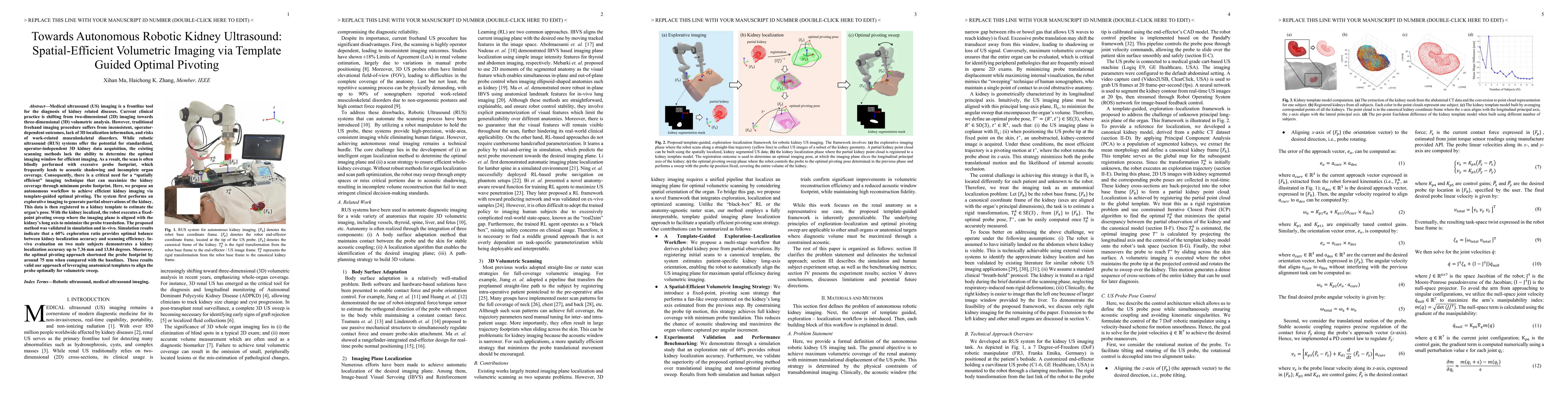
Medical ultrasound (US) imaging is a frontline tool for the diagnosis of kidney diseases. However, traditional freehand imaging procedure suffers from inconsistent, operator-dependent outcomes, lack of 3D localization information, and risks of work-related musculoskeletal disorders. While robotic ultrasound (RUS) systems offer the potential for standardized, operator-independent 3D kidney data acquisition, the existing scanning methods lack the ability to determine the optimal imaging window for efficient imaging. As a result, the scan is often blindly performed with excessive probe footprint, which frequently leads to acoustic shadowing and incomplete organ coverage. Consequently, there is a critical need for a spatially efficient imaging technique that can maximize the kidney coverage through minimum probe footprint. Here, we propose an autonomous workflow to achieve efficient kidney imaging via template-guided optimal pivoting. The system first performs an explorative imaging to generate partial observations of the kidney. This data is then registered to a kidney template to estimate the organ pose. With the kidney localized, the robot executes a fixed-point pivoting sweep where the imaging plane is aligned with the kidney long axis to minimize the probe translation. The proposed method was validated in simulation and in-vivo. Simulation results indicate that a 60% exploration ratio provides optimal balance between kidney localization accuracy and scanning efficiency. In-vivo evaluation on two male subjects demonstrates a kidney localization accuracy up to 7.36 mm and 13.84 degrees. Moreover, the optimal pivoting approach shortened the probe footprint by around 75 mm when compared with the baselines. These results valid our approach of leveraging anatomical templates to align the probe optimally for volumetric sweep.
Key Findings, in focus

Seven facets of this paper, analysed and brought into focus by AI.

AI-generated · verify against source Generated Feb 19, 2026
In focus · one-line synthesis

Provides a spatially efficient, operator-independent method for 3‑D kidney imaging, reducing probe footprint and improving coverage, which can enhance diagnostic accuracy and reduce clinician workload.

01
MethodologyHow they did it

Autonomous robotic kidney ultrasound workflow that performs exploratory imaging, registers partial kidney observations to a statistical kidney template using ICP with plane-to-plane cost and constrained rotation, computes an optimal pivoting pose aligning the probe’s long axis with the kidney’s longitudinal axis, and executes a fixed-point pivot sweep for volumetric coverage.

02
ResultsWhat they found
  • 60% exploration ratio yields stable localization with translation error ~10.8 mm and rotation error ~11.6°;
  • Simulation localization accuracy averages 8.73 mm translation and 12.86° rotation error;
  • Optimal pivoting reduces probe footprint by ~75 mm and imaging window displacement compared to baseline, achieving full coverage with fewer US frames.
03
SignificanceWhy it matters

Provides a spatially efficient, operator-independent method for 3‑D kidney imaging, reducing probe footprint and improving coverage, which can enhance diagnostic accuracy and reduce clinician workload.

04
Technical ContributionsWhat's new technically

Design of a template‑guided ICP registration with plane‑to‑plane cost and rotation clamping, and derivation of an optimal pivoting pose that aligns the probe’s long axis with the kidney’s longitudinal axis to minimize probe translation.

05
NoveltyWhat sets it apart

First autonomous robotic kidney ultrasound system that leverages a statistical kidney template for pose estimation and an optimal pivoting strategy to achieve efficient volumetric imaging with minimal probe footprint, combining exploration, registration, and pivot sweep in a unified workflow.

06
LimitationsWhat to be cautious about
  • Localization accuracy limited by inter‑patient anatomical variation and ultrasound segmentation noise;
  • Requires manual initial probe placement and patient breath‑hold, limiting fully autonomous operation.
07
Future WorkWhere it goes next
  • Integrate automatic probe placement and real‑time motion compensation;
  • Extend the framework to other abdominal organs and larger patient cohorts;
  • Incorporate acoustic modeling and tissue deformation for more realistic imaging.

Paper Details

Paper ID: 2602.16641
Categories:
cs.RO

How to Cite This Paper

Reference manager: RIS EndNote


Discussion 0

This website uses cookies to ensure you get the best experience. Learn more