This paper introduces a vision transformer-based model for cervical cell image classification, achieving an F1-score of 0.85227, slightly better than the baseline. It employs a four-step pipeline and uses Kernel SHAP for interpretability, highlighting key morphological and staining features.
We propose a novel approach to cervical cell image classification for cervical cancer screening using the EVA-02 transformer model. We developed a four-step pipeline: fine-tuning EVA-02, feature extraction, selecting important features through multiple machine learning models, and training a new artificial neural network with optional loss weighting for improved generalization. With this design, our best model achieved an F1-score of 0.85227, outperforming the baseline EVA-02 model (0.84878). We also utilized Kernel SHAP analysis and identified key features correlating with cell morphology and staining characteristics, providing interpretable insights into the decision-making process of the fine-tuned model. Our code is available at https://github.com/Khoa-NT/isbi2025_ps3c.
Generated Jun 08, 2025
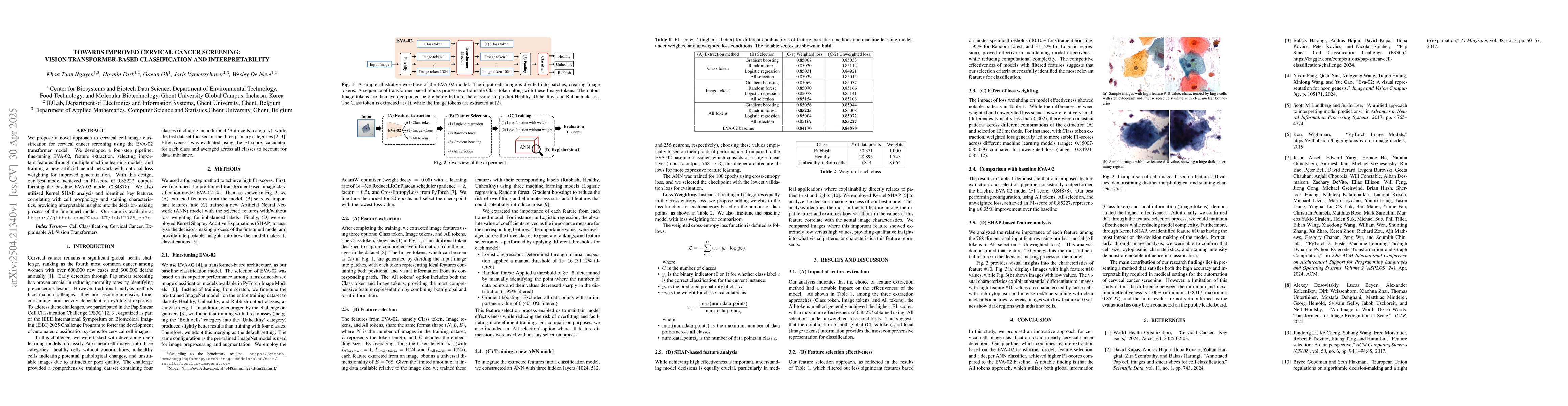
The research proposes a novel approach for cervical cell image classification using the EVA-02 transformer model, employing a four-step pipeline: fine-tuning EVA-02, feature extraction, selecting important features via multiple machine learning models, and training a new artificial neural network with optional loss weighting.
This research is important for improving cervical cancer screening accuracy by leveraging advanced transformer-based models and offering interpretability through SHAP analysis, which can aid in understanding model decisions and building trust in AI-driven diagnostic tools.
The paper introduces a transformer-based model (EVA-02) fine-tuned for cervical cell image classification, combined with feature selection and a custom neural network, resulting in improved classification performance.
This work stands out by applying transformer models to cervical cancer screening, emphasizing both classification accuracy and model interpretability through SHAP analysis, which is often overlooked in deep learning-based medical image analysis.
Current paper (gray), citations (green), references (blue)
Display is limited for performance on very large graphs.
Mayukha Pal, Asima Pradhan, Bhaswati Singha Deo et al.
Yao Qiang, Dongxiao Zhu, Prashant Khanduri et al.
Bishesh Khanal, Kanchan Poudel, Lisasha Poudel et al.
No citations found for this paper.
Comments (0)