Summary
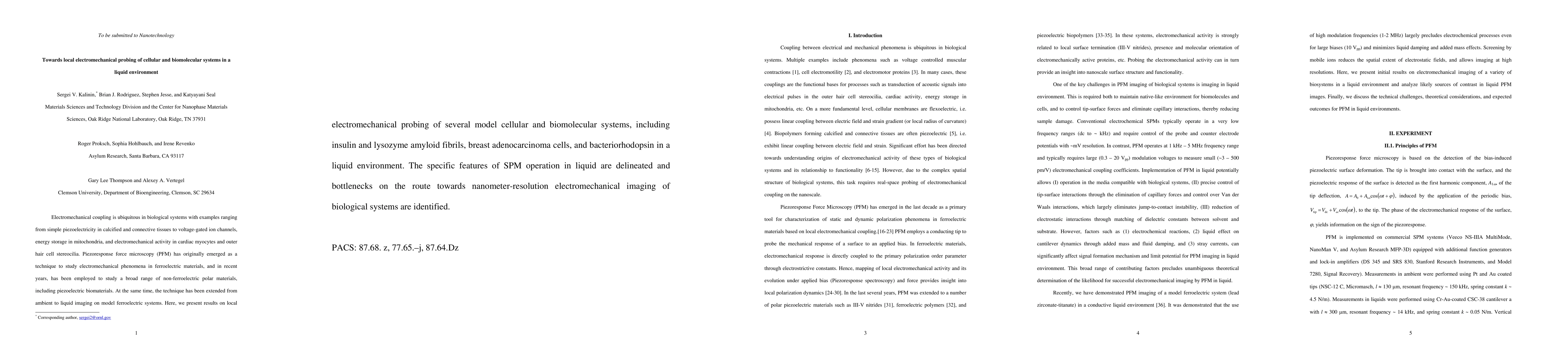
Electromechanical coupling is ubiquitous in biological systems with examples ranging from simple piezoelectricity in calcified and connective tissues to voltage-gated ion channels, energy storage in mitochondria, and electromechanical activity in cardiac myocytes and outer hair cell stereocilia. Piezoresponse force microscopy (PFM) has originally emerged as a technique to study electromechanical phenomena in ferroelectric materials, and in recent years, has been employed to study a broad range of non-ferroelectric polar materials, including piezoelectric biomaterials. At the same time, the technique has been extended from ambient to liquid imaging on model ferroelectric systems. Here, we present results on local electromechanical probing of several model cellular and biomolecular systems, including insulin and lysozyme amyloid fibrils, breast adenocarcinoma cells, and bacteriorhodopsin in a liquid environment. The specific features of SPM operation in liquid are delineated and bottlenecks on the route towards nanometer-resolution electromechanical imaging of biological systems are identified.
AI Key Findings
Get AI-generated insights about this paper's methodology, results, and significance.
Paper Details
PDF Preview
Key Terms
Citation Network
Current paper (gray), citations (green), references (blue)
Display is limited for performance on very large graphs.
Similar Papers
Found 4 papersCondensation and activator/repressor control of a transcription-regulated biomolecular liquid
Omar A. Saleh, Sam Wilken, Gabrielle R. Abraham
| Title | Authors | Year | Actions |
|---|
Comments (0)