Summary
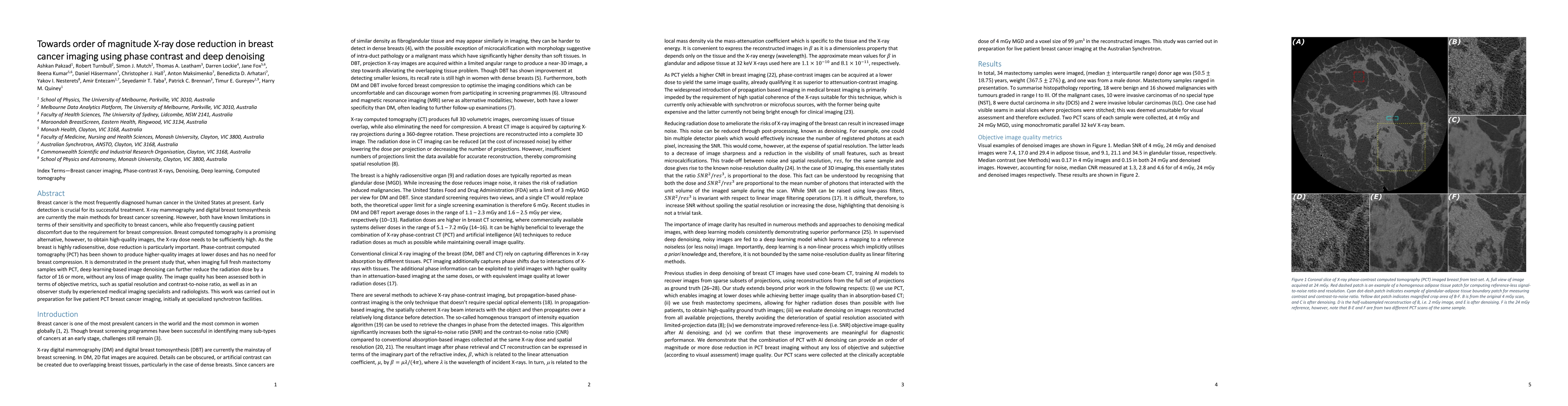
Breast cancer is the most frequently diagnosed human cancer in the United States at present. Early detection is crucial for its successful treatment. X-ray mammography and digital breast tomosynthesis are currently the main methods for breast cancer screening. However, both have known limitations in terms of their sensitivity and specificity to breast cancers, while also frequently causing patient discomfort due to the requirement for breast compression. Breast computed tomography is a promising alternative, however, to obtain high-quality images, the X-ray dose needs to be sufficiently high. As the breast is highly radiosensitive, dose reduction is particularly important. Phase-contrast computed tomography (PCT) has been shown to produce higher-quality images at lower doses and has no need for breast compression. It is demonstrated in the present study that, when imaging full fresh mastectomy samples with PCT, deep learning-based image denoising can further reduce the radiation dose by a factor of 16 or more, without any loss of image quality. The image quality has been assessed both in terms of objective metrics, such as spatial resolution and contrast-to-noise ratio, as well as in an observer study by experienced medical imaging specialists and radiologists. This work was carried out in preparation for live patient PCT breast cancer imaging, initially at specialized synchrotron facilities.
AI Key Findings
Generated Jun 08, 2025
Methodology
The research methodology involved imaging full fresh mastectomy samples with phase-contrast computed tomography (PCT) and applying deep learning-based image denoising techniques to reduce radiation doses.
Key Results
- Deep learning-based image denoising reduced radiation dose by a factor of 16 or more without loss of image quality when used with PCT.
- Image quality was assessed using objective metrics like spatial resolution and contrast-to-noise ratio, as well as through observer studies by medical imaging specialists and radiologists.
- The study demonstrates the potential for high-quality breast cancer imaging at significantly lower doses, paving the way for live patient PCT breast cancer imaging initially at specialized synchrotron facilities.
Significance
This research is crucial for breast cancer detection as it aims to reduce patient discomfort and radiation exposure associated with current imaging methods, while maintaining high diagnostic image quality.
Technical Contribution
The integration of phase-contrast computed tomography with deep learning-based image denoising for significant X-ray dose reduction in breast cancer imaging.
Novelty
This work stands out by combining phase-contrast imaging, known for its lower-dose potential, with deep learning denoising to achieve an order of magnitude reduction in radiation dose, maintaining high image quality.
Limitations
- The study was conducted using fresh mastectomy samples, so further research is needed to validate results in live patients.
- Access to specialized synchrotron facilities may limit immediate widespread application.
Future Work
- Transitioning from mastectomy samples to live patient PCT breast cancer imaging.
- Exploring the implementation of PCT with deep learning denoising at standard medical imaging facilities.
Paper Details
PDF Preview
Citation Network
Current paper (gray), citations (green), references (blue)
Display is limited for performance on very large graphs.
Similar Papers
Found 4 papersNo citations found for this paper.
Comments (0)