ArxivLens

Publication

Published: Jun 27, 2025
Updated: Aug 04, 2025
Categories: eess.IV, cs.CV

Metrics

Source: ArXiv

Quick Answers

What methodology did the authors use?

The study proposes a deep learning framework for white matter lesion (WMH) segmentation and localization using single- and multi-modal MRI inputs, directly in native space. It evaluates four input configurations: FLAIR-only, T1-only, concatenated FLAIR and T1, and modality-interchangeable setup. A multi-task model is introduced for jointly predicting lesion and anatomical region masks to estimate region-wise lesion burden. More in Methodology →

What are the key results?

Multimodal input significantly improves segmentation performance, outperforming unimodal models. — The modality-interchangeable setting trades accuracy for robustness, enabling inference in cases with missing modalities. More in Key Results →

Why is this work significant?

This research highlights the utility of multimodal fusion for accurate and robust WMH analysis, and the potential of joint modeling for integrated predictions, which can be beneficial for both clinical and research settings. More in Significance →

What are the main limitations?

Multi-task learning introduced a trade-off, with reduced multimodal performance compared to task-specific models. — The study did not extensively explore alternative training strategies to mitigate task interference. More in Limitations →

Paper Preview

Abstract

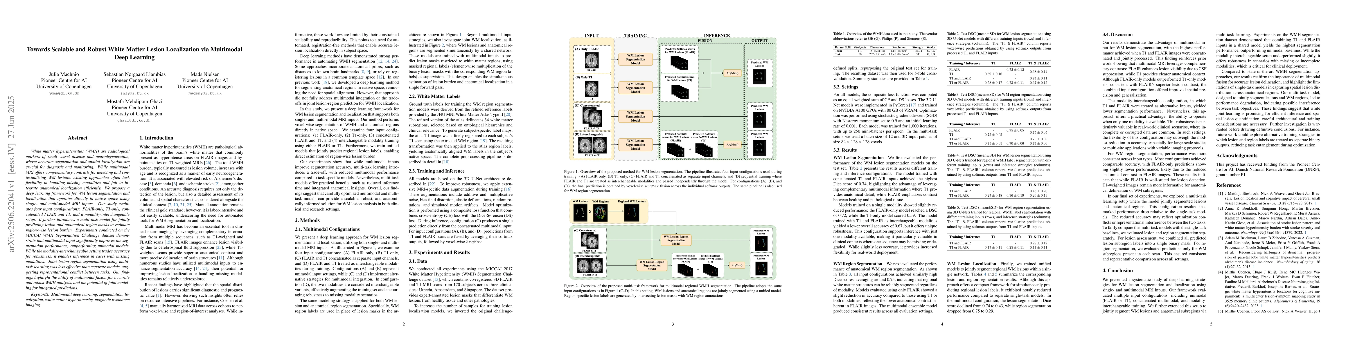
White matter hyperintensities (WMH) are radiological markers of small vessel disease and neurodegeneration, whose accurate segmentation and spatial localization are crucial for diagnosis and monitoring. While multimodal MRI offers complementary contrasts for detecting and contextualizing WM lesions, existing approaches often lack flexibility in handling missing modalities and fail to integrate anatomical localization efficiently. We propose a deep learning framework for WM lesion segmentation and localization that operates directly in native space using single- and multi-modal MRI inputs. Our study evaluates four input configurations: FLAIR-only, T1-only, concatenated FLAIR and T1, and a modality-interchangeable setup. It further introduces a multi-task model for jointly predicting lesion and anatomical region masks to estimate region-wise lesion burden. Experiments conducted on the MICCAI WMH Segmentation Challenge dataset demonstrate that multimodal input significantly improves the segmentation performance, outperforming unimodal models. While the modality-interchangeable setting trades accuracy for robustness, it enables inference in cases with missing modalities. Joint lesion-region segmentation using multi-task learning was less effective than separate models, suggesting representational conflict between tasks. Our findings highlight the utility of multimodal fusion for accurate and robust WMH analysis, and the potential of joint modeling for integrated predictions.

Key Findings, in focus

Seven facets of this paper, analysed and brought into focus by AI.

AI-generated · verify against source Generated Sep 07, 2025
In focus · one-line synthesis

This research highlights the utility of multimodal fusion for accurate and robust WMH analysis, and the potential of joint modeling for integrated predictions, which can be beneficial for both clinical and research settings.

01
MethodologyHow they did it

The study proposes a deep learning framework for white matter lesion (WMH) segmentation and localization using single- and multi-modal MRI inputs, directly in native space. It evaluates four input configurations: FLAIR-only, T1-only, concatenated FLAIR and T1, and modality-interchangeable setup. A multi-task model is introduced for jointly predicting lesion and anatomical region masks to estimate region-wise lesion burden.

02
ResultsWhat they found
  • Multimodal input significantly improves segmentation performance, outperforming unimodal models.
  • The modality-interchangeable setting trades accuracy for robustness, enabling inference in cases with missing modalities.
  • Joint lesion-region segmentation using multi-task learning was less effective than separate models, suggesting representational conflict between tasks.
03
SignificanceWhy it matters

This research highlights the utility of multimodal fusion for accurate and robust WMH analysis, and the potential of joint modeling for integrated predictions, which can be beneficial for both clinical and research settings.

04
Technical ContributionsWhat's new technically

The paper presents a deep learning framework for WMH segmentation and localization, evaluating multiple input configurations and introducing a multi-task model for joint lesion and anatomical region prediction.

05
NoveltyWhat sets it apart

The approach combines multimodal fusion with a multi-task learning strategy for WMH analysis, offering a scalable and robust solution for both clinical and research applications.

06
LimitationsWhat to be cautious about
  • Multi-task learning introduced a trade-off, with reduced multimodal performance compared to task-specific models.
  • The study did not extensively explore alternative training strategies to mitigate task interference.
07
Future WorkWhere it goes next
  • Investigate alternative training strategies where lesion and region labels are treated as separate binary outputs.
  • Explore the impact of incorporating additional MRI modalities or other imaging techniques.

Impact

26
References

Paper Details

Paper ID: 2506.22041
Comments: 2nd Sorbonne-Heidelberg Workshop on AI in medicine: Machine Learning for multi-modal data
Categories:
eess.IV cs.CV

How to Cite This Paper

Reference manager: RIS EndNote

Citation Network

Current paper (gray), citations (green), references (blue)

Display is limited for performance on very large graphs.

Current Paper
Citations
References
Click to view


Discussion 0

This website uses cookies to ensure you get the best experience. Learn more