Publication
Metrics
AI Quick Summary
This guide reviews the principles and variants of traction force microscopy on soft elastic substrates, focusing on strategies for reconstructing cellular forces amidst experimental noise. It discusses different approaches from elasticity theory and the role of biophysical models, along with practical aspects like substrate prep, image processing, and software availability.
Paper Preview
Abstract
The measurement of cellular traction forces on soft elastic substrates has become a standard tool for many labs working on mechanobiology. Here we review the basic principles and different variants of this approach. In general, the extraction of the substrate displacement field from image data and the reconstruction procedure for the forces are closely linked to each other and limited by the presence of experimental noise. We discuss different strategies to reconstruct cellular forces as they follow from the foundations of elasticity theory, including two- versus three-dimensional, inverse versus direct and linear versus non-linear approaches. We also discuss how biophysical models can improve force reconstruction and comment on practical issues like substrate preparation, image processing and the availability of software for traction force microscopy.
AI Key Findings
Get AI-generated insights about this paper's methodology, results, significance, and more — seven facets brought into focus.
Impact
Paper Details
PDF Preview
Key Terms
Citation Network
Current paper (gray), citations (green), references (blue)
Display is limited for performance on very large graphs.
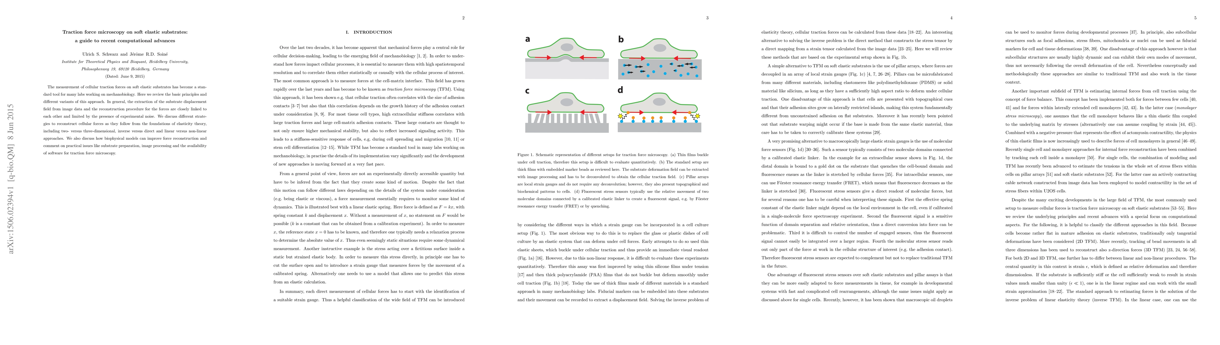
Discussion 0