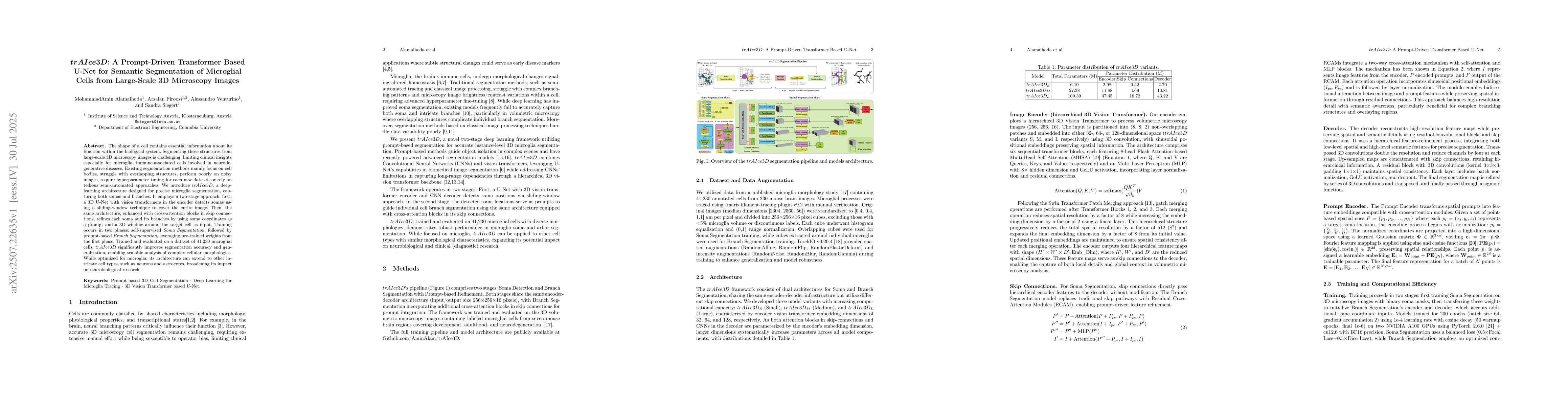
The shape of a cell contains essential information about its function within
the biological system. Segmenting these structures from large-scale 3D
microscopy images is challenging, limiting clinical insights especially for
microglia, immune-associated cells involved in neurodegenerative diseases.
Existing segmentation methods mainly focus on cell bodies, struggle with
overlapping structures, perform poorly on noisy images, require hyperparameter
tuning for each new dataset, or rely on tedious semi-automated approaches. We
introduce trAIce3D, a deep-learning architecture designed for precise microglia
segmentation, capturing both somas and branches. It employs a two-stage
approach: first, a 3D U-Net with vision transformers in the encoder detects
somas using a sliding-window technique to cover the entire image. Then, the
same architecture, enhanced with cross-attention blocks in skip connections,
refines each soma and its branches by using soma coordinates as a prompt and a
3D window around the target cell as input. Training occurs in two phases:
self-supervised Soma Segmentation, followed by prompt-based Branch
Segmentation, leveraging pre-trained weights from the first phase. Trained and
evaluated on a dataset of 41,230 microglial cells, trAIce3D significantly
improves segmentation accuracy and generalization, enabling scalable analysis
of complex cellular morphologies. While optimized for microglia, its
architecture can extend to other intricate cell types, such as neurons and
astrocytes, broadening its impact on neurobiological research.
Discussion 0