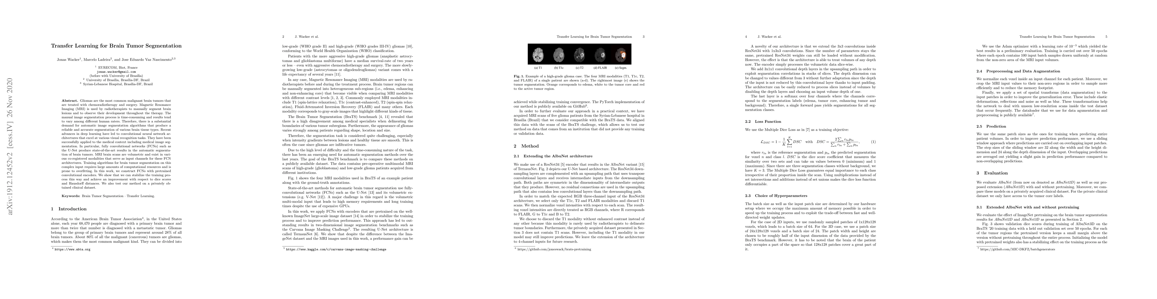
Publication
Metrics
AI Quick Summary
This paper explores the use of transfer learning with fully convolutional networks (FCNs) like U-Net to improve the automatic segmentation of brain tumors from MRI scans. By utilizing pretrained convolutional encoders, the authors stabilize training and achieve better segmentation accuracy, as demonstrated by improved dice scores and Hausdorff distances on a clinical dataset.
Paper Preview
Abstract
Gliomas are the most common malignant brain tumors that are treated with chemoradiotherapy and surgery. Magnetic Resonance Imaging (MRI) is used by radiotherapists to manually segment brain lesions and to observe their development throughout the therapy. The manual image segmentation process is time-consuming and results tend to vary among different human raters. Therefore, there is a substantial demand for automatic image segmentation algorithms that produce a reliable and accurate segmentation of various brain tissue types. Recent advances in deep learning have led to convolutional neural network architectures that excel at various visual recognition tasks. They have been successfully applied to the medical context including medical image segmentation. In particular, fully convolutional networks (FCNs) such as the U-Net produce state-of-the-art results in the automatic segmentation of brain tumors. MRI brain scans are volumetric and exist in various co-registered modalities that serve as input channels for these FCN architectures. Training algorithms for brain tumor segmentation on this complex input requires large amounts of computational resources and is prone to overfitting. In this work, we construct FCNs with pretrained convolutional encoders. We show that we can stabilize the training process this way and achieve an improvement with respect to dice scores and Hausdorff distances. We also test our method on a privately obtained clinical dataset.
AI Key Findings
Get AI-generated insights about this paper's methodology, results, significance, and more — seven facets brought into focus.
Impact
Paper Details
Authors
PDF Preview
Key Terms
Citation Network
Current paper (gray), citations (green), references (blue)
Display is limited for performance on very large graphs.
Discussion 0