Summary

This study evaluates publicly available deep-learning based lung segmentation models in transplant-eligible patients to determine their performance across disease severity levels, pathology categories, and lung sides, and to identify limitations impacting their use in preoperative planning in lung 
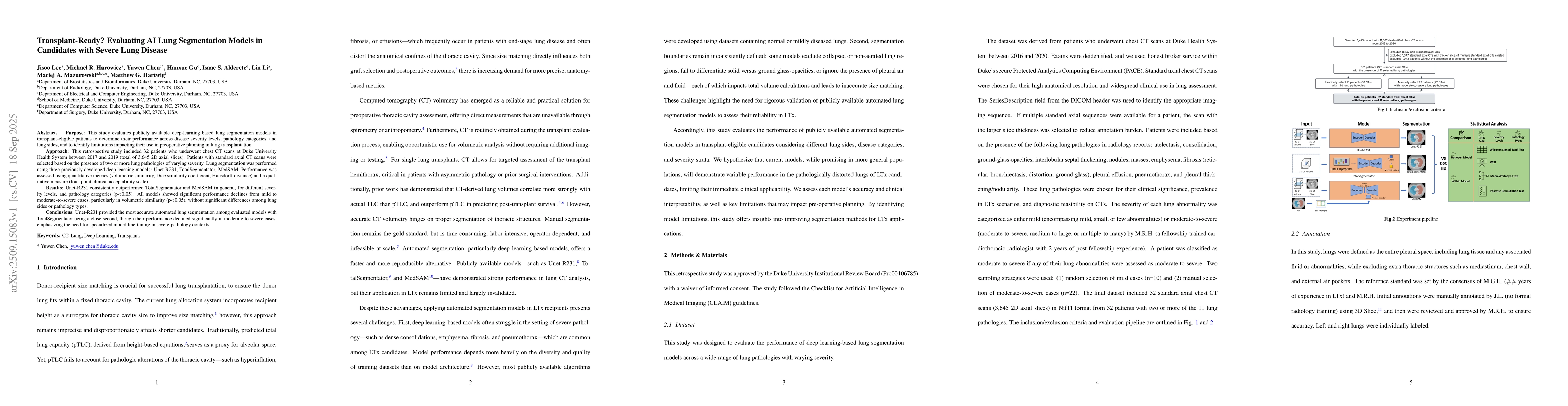
transplantation. This retrospective study included 32 patients who underwent chest CT scans at Duke University Health System between 2017 and 2019 (total of 3,645 2D axial slices). Patients with standard axial CT scans were selected based on the presence of two or more lung pathologies of varying severity. Lung segmentation was performed using three previously developed deep learning models: Unet-R231, TotalSegmentator, MedSAM. Performance was assessed using quantitative metrics (volumetric similarity, Dice similarity coefficient, Hausdorff distance) and a qualitative measure (four-point clinical acceptability scale). Unet-R231 consistently outperformed TotalSegmentator and MedSAM in general, for different severity levels, and pathology categories (p<0.05). All models showed significant performance declines from mild to moderate-to-severe cases, particularly in volumetric similarity (p<0.05), without significant differences among lung sides or pathology types. Unet-R231 provided the most accurate automated lung segmentation among evaluated models with TotalSegmentator being a close second, though their performance declined significantly in moderate-to-severe cases, emphasizing the need for specialized model fine-tuning in severe pathology contexts.

AI Key Findings

Generated Sep 30, 2025

Methodology

Retrospective study using 32 patients with severe lung disease, evaluated three AI models (Unet-R231, TotalSegmentator, MedSAM) via quantitative metrics (volumetric similarity, Dice similarity coefficient, Hausdorff distance) and qualitative clinical acceptability scale.

Key Results

  • Unet-R231 outperformed TotalSegmentator and MedSAM across all severity levels and pathology categories (p<0.05).
  • All models showed significant performance declines in moderate-to-severe cases, particularly in volumetric similarity (p<0.05).
  • Unet-R231 provided the most accurate segmentation, but all models exhibited systematic under-segmentation, leading to potential underestimation of thoracic cavity volume.

Significance

This research highlights the critical need for specialized AI models in lung transplant planning, as existing models struggle with severe lung pathologies, impacting donor-recipient size matching and surgical outcomes.

Technical Contribution

Comprehensive evaluation of AI lung segmentation models in transplant-eligible patients with severe lung disease using rigorous statistical methods and multiple performance metrics.

Novelty

First study to specifically evaluate AI models in the context of lung transplantation candidates with complex, severe lung pathologies, emphasizing clinical relevance for preoperative planning.

Limitations

  • Small sample size (32 patients) and imbalanced pathology distribution may limit generalizability.
  • CT scans were from a single institution, lacking control over imaging protocols and scanner types.

Future Work

  • Develop models fine-tuned on datasets enriched with moderate-to-severe lung pathologies.
  • Establish standardized segmentation protocols to avoid underestimation in volumetric analyses.

Paper Details

PDF Preview

Download PDF

Comments (0)

This website uses cookies to ensure you get the best experience. Learn more