Transport of Intensity Equation Microscopy for Dynamic Microtubules
Publication
Metrics
AI Quick Summary
This paper introduces Transport of Intensity Equation (TIE) Microscopy for imaging dynamic microtubules, demonstrating a three-fold improvement in signal-to-noise ratio over video-enhanced bright field imaging, and eliminating the need for frequent background updates.
Paper Preview
Abstract
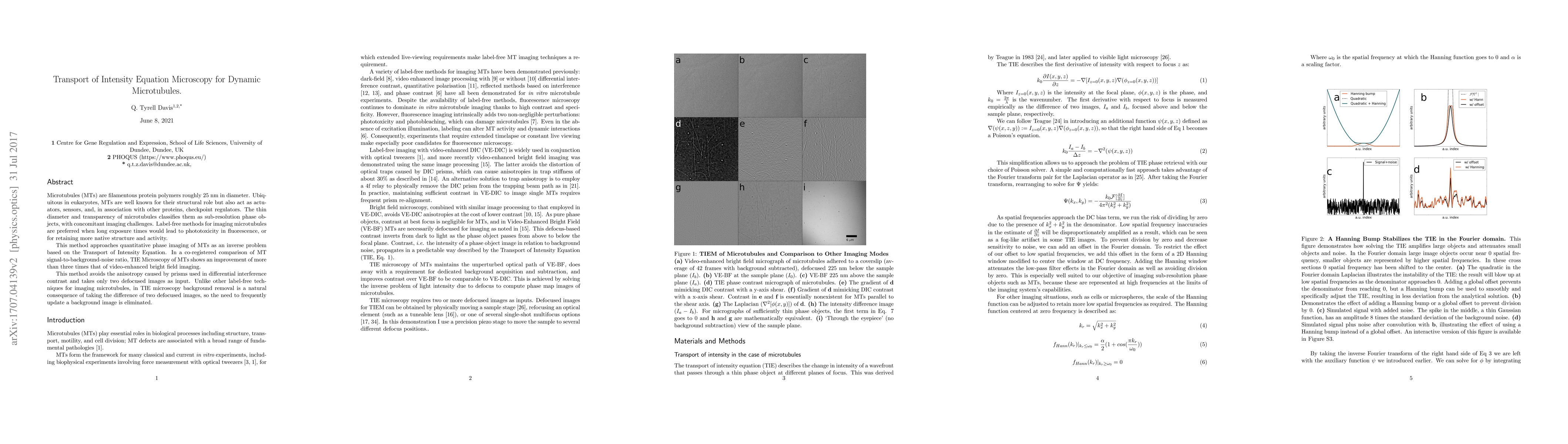
Microtubules (MTs) are filamentous protein polymers roughly 25 nm in diameter. Ubiquitous in eukaryotes, MTs are well known for their structural role but also act as actuators, sensors, and, in association with other proteins, checkpoint regulators. The thin diameter and transparency of microtubules classifies them as sub-resolution phase objects, with concomitant imaging challenges. Label-free methods for imaging microtubules are preferred when long exposure times would lead to phototoxicity in fluorescence, or for retaining more native structure and activity. This method approaches quantitative phase imaging of MTs as an inverse problem based on the Transport of Intensity Equation. In a co-registered comparison of MT signal-to-background-noise ratio, TIE Microscopy of MTs shows an improvement of more than three times that of video-enhanced bright field imaging. This method avoids the anisotropy caused by prisms used in differential interference contrast and takes only two defocused images as input. Unlike other label-free techniques for imaging microtubules, in TIE microscopy background removal is a natural consequence of taking the difference of two defocused images, so the need to frequently update a background image is eliminated.
AI Key Findings
Get AI-generated insights about this paper's methodology, results, significance, and more — seven facets brought into focus.
Impact
Paper Details
PDF Preview
Key Terms
Citation Network
Current paper (gray), citations (green), references (blue)
Display is limited for performance on very large graphs.
Discussion 0