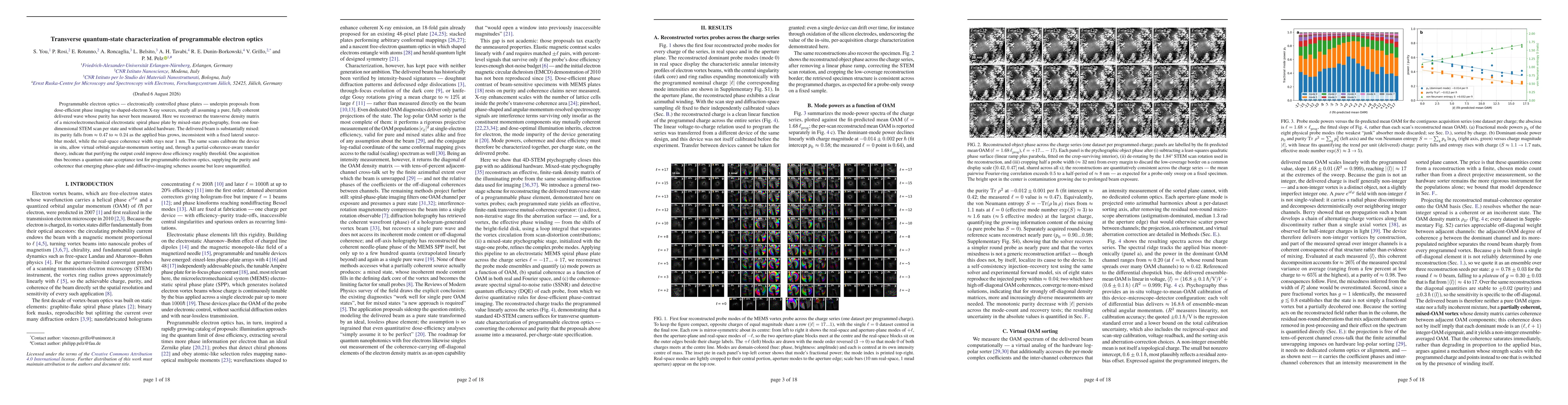
The study combines a two-stage reconstruction workflow using mixed-state ptychography on 4D-STEM data obtained from a programmable MEMS spiral phase plate. Stage one non-iteratively fits the aberration surface and vortex phase winding from bright-field shifts to initialize the probe. Stage two refines the complex probe modes via mixed-state ptychography to recover a finite-rank density matrix representing the transverse quantum state of the beam for each programmed OAM state (ℓ from −17 to +17).
Transverse quantum-state characterization of programmable electron optics
Publication
Metrics
Quick Answers
What methodology did the authors use?
The study combines a two-stage reconstruction workflow using mixed-state ptychography on 4D-STEM data obtained from a programmable MEMS spiral phase plate. Stage one non-iteratively fits the aberration surface and vortex phase winding from bright-field shifts to initialize the probe. Stage two refines the complex probe modes via mixed-state ptychography to recover a finite-rank density matrix representing the transverse quantum state... More in Methodology →
What are the key results?
The delivered beam is a mixed state with purity decreasing from ~0.47 to ~0.24 as the bias increases, inconsistent with a fixed lateral source-blur model. — Real-space coherence width remains near 1 nm across the OAM series, indicating preserved spatial coherence despite increasing impurity. More in Key Results →
Why is this work significant?
Provides the first direct, in-situ quantum-state characterization of programmable electron optics using 4D-STEM ptychography, delivering per-charge-state purity and coherence metrics that underpin dose-efficient phase imaging and shaped-electron sources. More in Significance →
What are the main limitations?
Finite-rank density matrix reconstruction may miss higher-order coherence components beyond the chosen model rank. — Results are device-specific to MEMS spiral phase plates and may not generalize to all programmable phase elements without adaptation. More in Limitations →
Abstract
Programmable electron optics -- electronically controlled phase plates -- underpin proposals from dose-efficient phase imaging to shaped-electron X-ray sources, nearly all assuming a pure, fully coherent delivered wave whose purity has never been measured. Here we reconstruct the transverse density matrix of a microelectromechanical electrostatic spiral phase plate by mixed-state ptychography, from one four-dimensional STEM scan per state and without added hardware. The delivered beam is substantially mixed: its purity falls from approximately 0.47 to approximately 0.24 as the applied bias grows, inconsistent with a fixed lateral source-blur model, while the real-space coherence width stays near 1 nm. The same scans calibrate the device in situ, allow virtual orbital-angular-momentum sorting and, through a partial-coherence-aware transfer theory, indicate that purifying the output could improve dose efficiency roughly threefold. One acquisition thus becomes a quantum-state acceptance test for programmable electron optics, supplying the purity and coherence that emerging phase-plate and diffractive-imaging schemes assume but leave unquantified.
Key Findings, in focus
Seven facets of this paper, analysed and brought into focus by AI.
Provides the first direct, in-situ quantum-state characterization of programmable electron optics using 4D-STEM ptychography, delivering per-charge-state purity and coherence metrics that underpin dose-efficient phase imaging and shaped-electron sources.
- The delivered beam is a mixed state with purity decreasing from ~0.47 to ~0.24 as the bias increases, inconsistent with a fixed lateral source-blur model.
- Real-space coherence width remains near 1 nm across the OAM series, indicating preserved spatial coherence despite increasing impurity.
- Virtual orbital-angular-momentum sorting and a partial-coherence-aware transfer theory quantify potential dose-efficiency improvements (~3x) if output purity is purified.
Provides the first direct, in-situ quantum-state characterization of programmable electron optics using 4D-STEM ptychography, delivering per-charge-state purity and coherence metrics that underpin dose-efficient phase imaging and shaped-electron sources.
Introduces a general two-stage mixed-state ptychography pipeline that reconstructs the transverse density matrix of a programmable electron-optics device from a single 4D-STEM scan per charge state, enabling in situ calibration and coherence-aware performance metrics.
First experimental report to reconstruct and quantify the full transverse quantum state (including off-diagonal coherences) of a programmable electron-optics element across multiple OAM states, revealing substantial mode impurity and its impact on dose efficiency.
- Finite-rank density matrix reconstruction may miss higher-order coherence components beyond the chosen model rank.
- Results are device-specific to MEMS spiral phase plates and may not generalize to all programmable phase elements without adaptation.
- Assumes accurate stage-one calibration; residual scan-distortion or aberration errors could bias the mixed-state reconstruction.
- Extend the methodology to other programmable phase elements (e.g., multi-electrode plates) to map broader coherence landscapes.
- Experimentally pursue hardware or operational strategies to increase output purity and thus dose efficiency across bias ranges.
- Incorporate full off-diagonal coherence characterization into real-time diagnostics for dynamic beam shaping.
Discussion 0