TriadNet: Sampling-free predictive intervals for lesional volume in 3D brain MR images
Publication
Metrics
AI Quick Summary
TriadNet proposes a multi-head CNN architecture for simultaneous segmentation and predictive interval estimation of brain lesion volumes in 3D MR images, enhancing clinical utility and decision-making. The method outperforms existing techniques on the BraTS 2021 dataset, providing results in under a second.
Paper Preview
Abstract
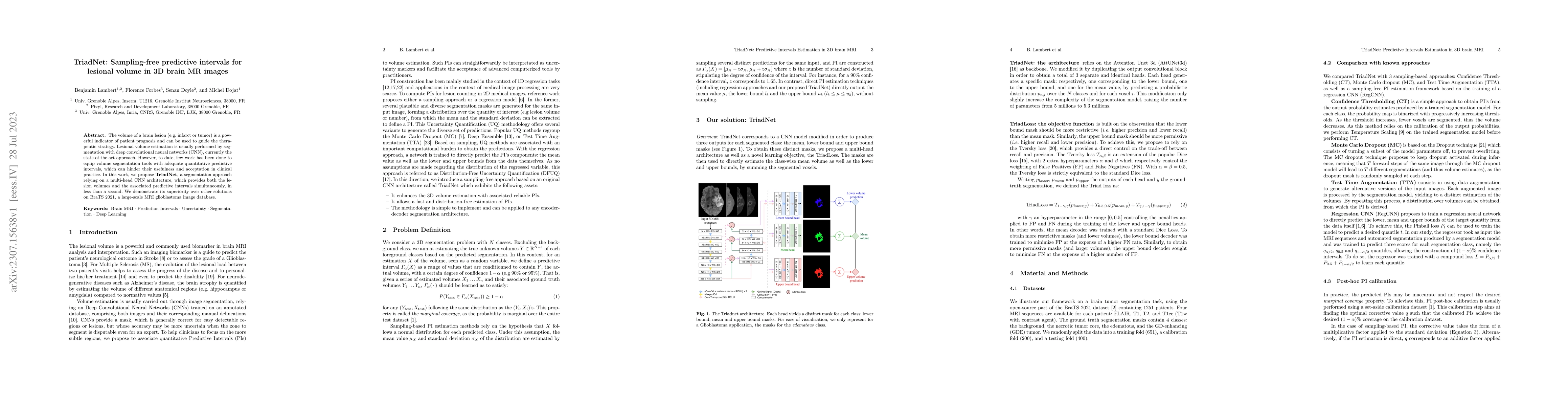
The volume of a brain lesion (e.g. infarct or tumor) is a powerful indicator of patient prognosis and can be used to guide the therapeutic strategy. Lesional volume estimation is usually performed by segmentation with deep convolutional neural networks (CNN), currently the state-of-the-art approach. However, to date, few work has been done to equip volume segmentation tools with adequate quantitative predictive intervals, which can hinder their usefulness and acceptation in clinical practice. In this work, we propose TriadNet, a segmentation approach relying on a multi-head CNN architecture, which provides both the lesion volumes and the associated predictive intervals simultaneously, in less than a second. We demonstrate its superiority over other solutions on BraTS 2021, a large-scale MRI glioblastoma image database.
AI Key Findings
Get AI-generated insights about this paper's methodology, results, significance, and more — seven facets brought into focus.
Impact
Paper Details
Authors
PDF Preview
Key Terms
Citation Network
Current paper (gray), citations (green), references (blue)
Display is limited for performance on very large graphs.
Discussion 0