The paper introduces CARP3D, a deep learning method that employs 2.5D multiple-instance learning to triage high-risk 2D slices from 3D pathology datasets, facilitating more efficient pathologist assessments. The approach enhances diagnostic accuracy for prostate cancer by integrating contextual depth information, achieving an AUC of 90.4% compared to 81.3% for independent 2D slice analysis.
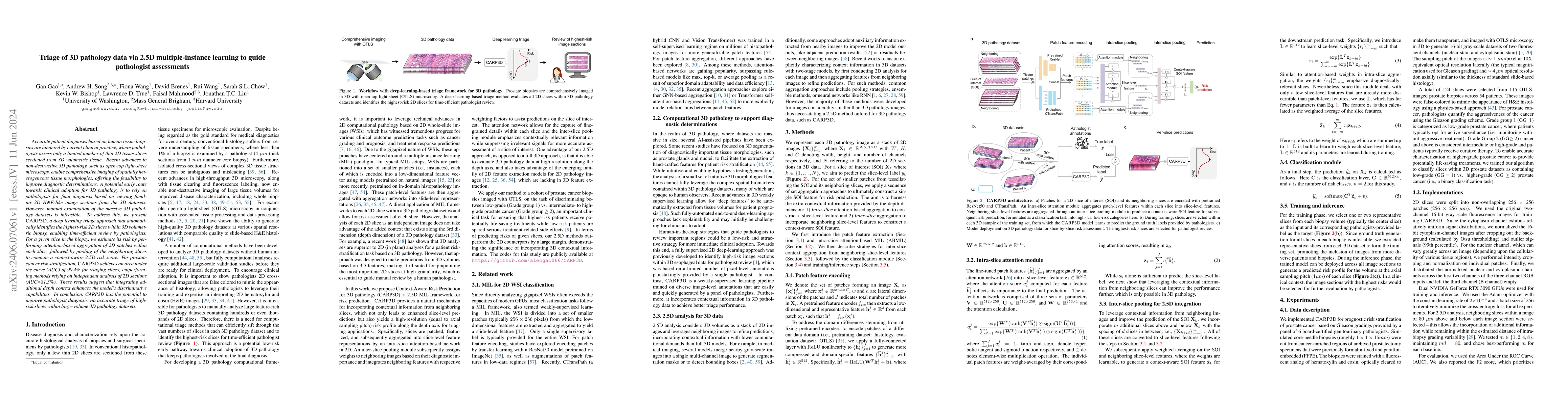
Accurate patient diagnoses based on human tissue biopsies are hindered by current clinical practice, where pathologists assess only a limited number of thin 2D tissue slices sectioned from 3D volumetric tissue. Recent advances in non-destructive 3D pathology, such as open-top light-sheet microscopy, enable comprehensive imaging of spatially heterogeneous tissue morphologies, offering the feasibility to improve diagnostic determinations. A potential early route towards clinical adoption for 3D pathology is to rely on pathologists for final diagnosis based on viewing familiar 2D H&E-like image sections from the 3D datasets. However, manual examination of the massive 3D pathology datasets is infeasible. To address this, we present CARP3D, a deep learning triage approach that automatically identifies the highest-risk 2D slices within 3D volumetric biopsy, enabling time-efficient review by pathologists. For a given slice in the biopsy, we estimate its risk by performing attention-based aggregation of 2D patches within each slice, followed by pooling of the neighboring slices to compute a context-aware 2.5D risk score. For prostate cancer risk stratification, CARP3D achieves an area under the curve (AUC) of 90.4% for triaging slices, outperforming methods relying on independent analysis of 2D sections (AUC=81.3%). These results suggest that integrating additional depth context enhances the model's discriminative capabilities. In conclusion, CARP3D has the potential to improve pathologist diagnosis via accurate triage of high-risk slices within large-volume 3D pathology datasets.
Get AI-generated insights about this paper's methodology, results, and significance.
Current paper (gray), citations (green), references (blue)
Chintan Shah, Harshith Padigela, John Abel et al.
Hassan Keshvarikhojasteh, Mitko Veta, Josien P. W. Pluim et al.
Stergios Christodoulidis, Maria Vakalopoulou, Paul-Henry Cournède et al.
No citations found for this paper.
Title | Authors | Year | Actions |
---|---|---|---|
Analysis of 3D pathology samples using weakly supervised AI
|
Andrew H. Song, Mane Williams, Drew F. K. Williamson, Sarah S. L. Chow, Guillaume Jaume, Gan Gao, Andrew Zhang, Bowen Chen, Alexander S. Baras, R. Serafin, Richard Colling, Michelle R. Downes, Xavier Farré, Peter Humphrey, Clare Verrill, Lawrence D. True, Anil V. Parwani, Jonathan T. C. Liu, Faisal Mahmood
|
2024
|
View Paper |
The 3D Revolution in Cancer Discovery.
|
Linghua Wang, Mingyao Li, Tae Hyun Hwang
|
2024
|
View Paper |
Multiscale Optical Imaging Fusion for Cervical Precancer Diagnosis: Integrating Widefield Colposcopy and High-Resolution Endomicroscopy
|
David Brenes, M. Salcedo, Jackson B. Coole, Yajur Maker, Alex Kortum, R. Schwarz, J. Carns, Imran S Vohra, J. C. Possati-Resende, Márcio Antoniazzi, Bruno de Oliveira Fonseca, Karen C Borba Souza, Iara V Vidigal Santana, Flávia Fazzio Barbin, Regis Kreitchmann, N. Ramanujam, Kathleen M. Schmeler, R. Richards-Kortum
|
2024
|
View Paper |
Towards a general-purpose foundation model for computational pathology.
|
Richard J. Chen, Tong Ding, Ming Y. Lu, Drew F. K. Williamson, Guillaume Jaume, Andrew H. Song, Bowen Chen, Andrew Zhang, Daniel Shao, Muhammad Shaban, Mane Williams, Lukas Oldenburg, Luca L. Weishaupt, Judy J. Wang, Anurag Vaidya, L. Le, Georg K. Gerber, S. Sahai, Walt Williams, Faisal Mahmood
|
2024
|
View Paper |
A visual-language foundation model for computational pathology.
|
Ming Y. Lu, Bowen Chen, Drew F. K. Williamson, Richard J. Chen, Ivy Liang, Tong Ding, Guillaume Jaume, Igor Odintsov, L. Le, Georg K. Gerber, Anil V. Parwani, Andrew Zhang, Faisal Mahmood
|
2024
|
View Paper |
An end-to-end workflow for nondestructive 3D pathology.
|
K. Bishop, Lindsey A. Erion Barner, Qinghua Han, Elena Baraznenok, Lydia Lan, Chetan Poudel, Gan Gao, R. Serafin, Sarah S. L. Chow, A. Glaser, Andrew Janowczyk, David Brenes, Hongyi Huang, Dominie Miyasato, Lawrence D. True, Soyoung Kang, J. Vaughan, Jonathan T. C. Liu
|
2024
|
View Paper |
Engineering the future of 3D pathology
|
Jonathan T C Liu, Sarah S. L. Chow, Richard Colling, M. Downes, Xavier Farré, Peter Humphrey, Andrew Janowczyk, T. Mirtti, Clare Verrill, Inti Zlobec, Lawrence D. True
|
2023
|
View Paper |
Artificial intelligence for digital and computational pathology
|
Andrew H. Song, Guillaume Jaume, Drew F. K. Williamson, Ming Y. Lu, Anurag Vaidya, Tiffany R. Miller, Faisal Mahmood
|
2023
|
View Paper |
A visual–language foundation model for pathology image analysis using medical Twitter
|
Zhi Huang, Federico Bianchi, Mert Yuksekgonul, T. Montine, J. Zou
|
2023
|
View Paper |
Transformer-based biomarker prediction from colorectal cancer histology: A large-scale multicentric study
|
S. Wagner, Daniel Reisenbüchler, N. West, J. Niehues, Jiefu Zhu, S. Foersch, G. P. Veldhuizen, P. Quirke, H. Grabsch, P. A. van den Brandt, G. Hutchins, S. Richman, T. Yuan, R. Langer, J. Jenniskens, K. Offermans, W. Mueller, R. Gray, S. Gruber, J. Greenson, G. Rennert, J. Bonner, Daniel Schmolze, J. Jonnagaddala, Nicholas J Hawkins, R. Ward, D. Morton, M. Seymour, L. Magill, M. Nowak, Jennifer Hay, V. Koelzer, D. Church, C. Matek, C. Geppert, Chaolong Peng, Cheng Zhi, Xiaoming Ouyang, J. James, M. Loughrey, M. Salto‐Tellez, H. Brenner, M. Hoffmeister, D. Truhn, J. Schnabel, M. Boxberg, Tingying Peng, Jakob Nikolas Kather, D. Church, E. Domingo, J. Edwards, B. Glimelius, I. Gøgenur, A. Harkin, Jennifer Hay, T. Iveson, E. Jaeger, C. Kelly, R. Kerr, N. Maka, Hannah Morgan, K. Oien, C. Orange, C. Palles, C. Roxburgh, O. Sansom, Mark P. Saunders, I. Tomlinson
|
2023
|
View Paper |
AI-triaged 3D pathology to improve detection of esophageal neoplasia while reducing pathologist workloads.
|
Lindsey A. Erion Barner, Gan Gao, Deepti M Reddi, Lydia Lan, Wynn Burke, F. Mahmood, W. Grady, Jonathan T. C. Liu
|
2023
|
View Paper |
Nondestructive 3D pathology with analysis of nuclear features for prostate cancer risk assessment
|
R. Serafin, C. Koyuncu, Weisi Xie, Hongyi Huang, A. Glaser, N. Reder, A. Janowczyk, L. True, A. Madabhushi, Jonathan T. C. Liu
|
2023
|
View Paper |
Video Pretraining Advances 3D Deep Learning on Chest CT Tasks
|
Alexander Ke, Shih-Cheng Huang, Chloe O'Connell, M. Klimont, Serena Yeung, P. Rajpurkar
|
2023
|
View Paper |
Detecting adrenal lesions on 3D CT scans using a 2.5D deep learning model
|
Sanson T. S. Poon, Fahmy W. F. Hanna, Fran¸cois Lemarchand, Cherian George, Alexander Clark, S. Lea, Charlie Coleman, Giuseppe Sollazzo
|
2023
|
View Paper |
Comprehensive Surface Histology of Fresh Resection Margins With Rapid Open-Top Light-Sheet (OTLS) Microscopy
|
Gan Gao, Dominie Miyasato, L. Barner, R. Serafin, K. Bishop, Weisi Xie, A. Glaser, E. Rosenthal, L. True, Jonathan T. C. Liu
|
2023
|
View Paper |
Nondestructive 3D Pathology Image Atlas of Barrett Esophagus With Open-Top Light-Sheet Microscopy.
|
Deepti M Reddi, L. Barner, Wynn Burke, Gan Gao, W. Grady, Jonathan T. C. Liu
|
2023
|
View Paper |
Comparing 3D, 2.5D, and 2D Approaches to Brain Image Auto-Segmentation
|
A. Avesta, Sajid Hossain, Mingde Lin, M. Aboian, H. Krumholz, Sanjay Aneja
|
2022
|
View Paper |
Embedding Space Augmentation for Weakly Supervised Learning in Whole-Slide Images
|
Imaad Zaffar, Guillaume Jaume, N. Rajpoot, Faisal Mahmood
|
2022
|
View Paper |
Derivation of prognostic contextual histopathological features from whole-slide images of tumours via graph deep learning.
|
Yongju Lee, Jeong Hwan Park, Sohee Oh, Kyoungseob Shin, Jiyu Sun, Minsun Jung, Cheol Lee, Hyojin Kim, Jin-Haeng Chung, K. Moon, Sung-Jun Kwon
|
2022
|
View Paper |
Transformer-based unsupervised contrastive learning for histopathological image classification
|
Xiyue Wang, Sen Yang, Jun Zhang, Minghui Wang, Jing Zhang, Wei Yang, Junzhou Huang, Xiao Han
|
2022
|
View Paper |
Incorporating intratumoral heterogeneity into weakly-supervised deep learning models via variance pooling
|
Iain Carmichael, Andrew H. Song, Richard J. Chen, Drew F. K. Williamson, Tiffany Y. Chen, Faisal Mahmood
|
2022
|
View Paper |
Scaling Vision Transformers to Gigapixel Images via Hierarchical Self-Supervised Learning
|
Richard J. Chen, Chengkuan Chen, Yicong Li, Tiffany Y. Chen, A. Trister, R. G. Krishnan, Faisal Mahmood
|
2022
|
View Paper |
A hybrid open-top light-sheet microscope for versatile multi-scale imaging of cleared tissues
|
A. Glaser, K. Bishop, L. Barner, E. Susaki, Shimpei I. Kubota, Gan Gao, R. Serafin, P. Balaram, Emily Turschak, P. Nicovich, H. Lai, L. Lucas, Yating Yi, Eva K. Nichols, Hongyi Huang, N. Reder, Jasmine Wilson, R. Sivakumar, E. Shamskhou, C. Stoltzfus, Xing Wei, Andrew K. Hempton, M. Pende, P. Murawala, H. Dodt, T. Imaizumi, J. Shendure, Brian J. Beliveau, Michael Y. Gerner, Li Xin, Hu Zhao, L. True, R. Reid, J. Chandrashekar, H. Ueda, K. Svoboda, Jonathan T. C. Liu
|
2022
|
View Paper |
Benchmarking weakly-supervised deep learning pipelines for whole slide classification in computational pathology
|
Narmin Ghaffari Laleh, H. Muti, C. Loeffler, A. Echle, O. Saldanha, Faisal Mahmood, Ming Y. Lu, Christian Trautwein, R. Langer, B. Dislich, R. D. Bülow, H. Grabsch, H. Brenner, J. Chang-Claude, E. Alwers, T. Brinker, Firas Khader, D. Truhn, N. Gaisa, P. Boor, M. Hoffmeister, V. Schulz, Jakob Nikolas Kather
|
2022
|
View Paper |
Deep learning-enabled assessment of cardiac allograft rejection from endomyocardial biopsies
|
Jana Lipková, Tiffany Y. Chen, Ming Y. Lu, Richard J. Chen, Maha Shady, Mane Williams, Jingwen Wang, Zahra Noor, R. Mitchell, Mehmet Turan, Gulfize Coskun, F. Yilmaz, D. Demir, D. Nart, K. Başak, N. Turhan, S. Ozkara, Y. Banz, K. Odening, Faisal Mahmood
|
2022
|
View Paper |
Artificial intelligence for diagnosis and Gleason grading of prostate cancer: the PANDA challenge
|
W. Bulten, K. Kartasalo, Po-Hsuan Cameron Chen, P. Ström, H. Pinckaers, K. Nagpal, Yuannan Cai, David F. Steiner, H. V. van Boven, Robert Vink, C. Hulsbergen–van de Kaa, J. A. van der Laak, M. Amin, A. Evans, T. H. van der Kwast, Robert Allan, P. Humphrey, H. Grönberg, H. Samaratunga, B. Delahunt, T. Tsuzuki, T. Häkkinen, L. Egevad, Maggie Demkin, Sohier Dane, Fraser Tan, Masi Valkonen, G. Corrado, L. Peng, C. Mermel, P. Ruusuvuori, G. Litjens, M. Eklund, Américo Aslı Xavier Katerina Vincent Guilherme Paromita Gü Brilhante Çakır Farré Geronatsiou Molinié Pereira , A. Brilhante, A. Çakır, X. Farré, K. Geronatsiou, V. Molinie, Guilherme Pereira, Paromita Roy, G. Saile, P. G. O. Salles, E. Schaafsma, J. Tschui, Jorge Billoch-Lima, Emíio M. Pereira, M. Zhou, Shujun He, Sejun Song, Qing Sun, H. Yoshihara, Taiki Yamaguchi, K. Ono, Tao Shen, Jianyi Ji, Arnaud Roussel, Kairong Zhou, Tianrui Chai, Nina Weng, Dmitry A. Grechka, M. Shugaev, Raphael Kiminya, V. Kovalev, D. Voynov, V. Malyshev, E. Lapo, Manolo Quispe Campos, Noriaki Ota, Shinsuke Yamaoka, Yusuke Fujimoto, Kentaro Yoshioka, Joni Juvonen, M. Tukiainen, A. Karlsson, Rui Guo, Chia-Lun Hsieh, I. Zubarev, Habib S. T. Bukhar, Wenyuan Li, Jiayun Li, W. Speier, C. Arnold, Kyungdoc Kim, Byeonguk Bae, Y. Kim, Hong-Seok Lee, Jeonghyuk Park
|
2022
|
View Paper |
Prostate Cancer Risk Stratification via Nondestructive 3D Pathology with Deep Learning–Assisted Gland Analysis
|
Weisi Xie, N. Reder, C. Koyuncu, P. Leo, S. Hawley, Hongyi Huang, C. Mao, N. Postupna, Soyoung Kang, R. Serafin, Gan Gao, Qinghua Han, K. Bishop, L. Barner, P. Fu, J. Wright, C. Keene, J. Vaughan, A. Janowczyk, A. Glaser, A. Madabhushi, L. True, Jonathan T. C. Liu
|
2021
|
View Paper |
Deep learning trained on hematoxylin and eosin tumor region of Interest predicts HER2 status and trastuzumab treatment response in HER2+ breast cancer
|
Saman Farahmand, Aileen I. Fernandez, F. Ahmed, D. Rimm, Jeffrey H. Chuang, Emily S. Reisenbichler, Kourosh Zarringhalam
|
2021
|
View Paper |
Whole Slide Images are 2D Point Clouds: Context-Aware Survival Prediction using Patch-based Graph Convolutional Networks
|
Richard J. Chen, Ming Y. Lu, M. Shaban, Chengkuan Chen, Tiffany Y. Chen, Drew F. K. Williamson, Faisal Mahmood
|
2021
|
View Paper |
TransMIL: Transformer based Correlated Multiple Instance Learning for Whole Slide Image Classication
|
Zhucheng Shao, Hao Bian, Yang Chen, Yifeng Wang, Jian Zhang, Xiangyang Ji, Yongbing Zhang
|
2021
|
View Paper |
Harnessing non-destructive 3D pathology
|
Jonathan T. C. Liu, A. Glaser, K. Bera, L. True, N. Reder, K. Eliceiri, A. Madabhushi
|
2021
|
View Paper |
Semi-automatic sigmoid colon segmentation in CT for radiation therapy treatment planning via an iterative 2.5-D deep learning approach
|
Y. Gonzalez, Chenyang Shen, Hyunuk Jung, D. Nguyen, Steve B. Jiang, K. Albuquerque, X. Jia
|
2020
|
View Paper |
Inter-Slice Context Residual Learning for 3D Medical Image Segmentation
|
Jianpeng Zhang, Yutong Xie, Yan Wang, Yong Xia
|
2020
|
View Paper |
3D virtual histology of human pancreatic tissue by multiscale phase-contrast X-ray tomography
|
J. Frohn, Diana Pinkert-Leetsch, Jeannine Missbach-Güntner, Marius Reichardt, M. Osterhoff, F. Alves, T. Salditt
|
2020
|
View Paper |
Multi-resolution open-top light-sheet microscopy to enable efficient 3D pathology workflows.
|
L. Barner, A. Glaser, Hongyi Huang, L. True, Jonathan T. C. Liu
|
2020
|
View Paper |
Bridging 2D and 3D segmentation networks for computation-efficient volumetric medical image segmentation: An empirical study of 2.5D solutions
|
Yichi Zhang, Qingcheng Liao, Leiting Ding, Jicong Zhang
|
2020
|
View Paper |
BRR-Net: A Tandem Architectural CNN-RNN for Automatic Body Region Localization in CT Images.
|
V. Agrawal, J. Udupa, Yubing Tong, D. Torigian
|
2020
|
View Paper |
FalseColor-Python: A rapid intensity-leveling and digital-staining package for fluorescence-based slide-free digital pathology
|
R. Serafin, Weisi Xie, A. Glaser, Jonathan T. C. Liu
|
2020
|
View Paper |
Data-efficient and weakly supervised computational pathology on whole-slide images
|
Ming Y. Lu, Drew F. K. Williamson, Tiffany Y. Chen, Richard J. Chen, Matteo Barbieri, Faisal Mahmood
|
2020
|
View Paper |
Deep learning for prediction of colorectal cancer outcome: a discovery and validation study
|
O. Skrede, S. D. Raedt, A. Kleppe, T. Hveem, K. Liestøl, J. Maddison, H. Askautrud, M. Pradhan, J. A. Nesheim, F. Albregtsen, I. Farstad, E. Domingo, D. Church, A. Nesbakken, N. Shepherd, I. Tomlinson, R. Kerr, M. Novelli, D. Kerr, H. Danielsen
|
2020
|
View Paper |
Three-dimensional visualization of cleared human pancreas cancer reveals that sustained epithelial-to-mesenchymal transition is not required for venous invasion
|
Seung‐Mo Hong, Dongjun Jung, A. Kiemen, M. Gaida, T. Yoshizawa, Alicia M. Braxton, M. Noë, Gemma Lionheart, K. Oshima, E. Thompson, R. Burkhart, Pei-Hsun Wu, D. Wirtz, R. Hruban, L. Wood
|
2019
|
View Paper |
Deep learning-based classification of mesothelioma improves prediction of patient outcome
|
P. Courtiol, C. Maussion, Matahi Moarii, E. Pronier, Samuel Pilcer, M. Sefta, Pierre Manceron, Sylvain Toldo, M. Zaslavskiy, N. Le Stang, N. Girard, O. Elemento, A. Nicholson, J. Blay, F. Galateau-Salle, G. Wainrib, T. Clozel
|
2019
|
View Paper |
A Cascade of 2.5D CNN and Bidirectional CLSTM Network for Mitotic Cell Detection in 4D Microscopy Image
|
Titinunt Kitrungrotsakul, Xianhua Han, Yutaro Iwamoto, S. Takemoto, H. Yokota, Sari Ipponjima, T. Nemoto, W. Xiong, Yenwei Chen
|
2019
|
View Paper |
Open-Top Light-Sheet Microscopy Image Atlas of Prostate Core Needle Biopsies.
|
N. Reder, A. Glaser, Erin McCarty, Ye Chen, L. True, Jonathan T. C. Liu
|
2019
|
View Paper |
Multi-immersion open-top light-sheet microscope for high-throughput imaging of cleared tissues
|
A. Glaser, N. Reder, Ye Chen, Chengbo Yin, Linpeng Wei, Soyoung Kang, L. Barner, Weisi Xie, Erin McCarty, C. Mao, Aaron R. Halpern, C. Stoltzfus, J. Daniels, Michael Y. Gerner, P. Nicovich, J. Vaughan, L. True, Jonathan T. C. Liu
|
2019
|
View Paper |
Classification and mutation prediction from non–small cell lung cancer histopathology images using deep learning
|
N. Coudray, P. Ocampo, T. Sakellaropoulos, N. Narula, M. Snuderl, D. Fenyö, A. Moreira, Narges Razavian, A. Tsirigos
|
2018
|
View Paper |
Deep CNNs for microscopic image classification by exploiting transfer learning and feature concatenation
|
Long D. Nguyen, Dongyun Lin, Zhiping Lin, Jiuwen Cao
|
2018
|
View Paper |
Diagnostic Efficacy of Deeper Sections in Routine Oral Histopathology Practice: a Retrospective Study
|
A. Yadav, M. Kamboj, A. Narwal, A. Devi
|
2018
|
View Paper |
Attention-based Deep Multiple Instance Learning
|
Maximilian Ilse, J. Tomczak, M. Welling
|
2018
|
View Paper |
Whole-tissue biopsy phenotyping of three-dimensional tumours reveals patterns of cancer heterogeneity
|
N. Tanaka, Shigeaki Kanatani, Raju Tomer, C. Sahlgren, P. Kronqvist, D. Kaczynska, L. Louhivuori, L. Kis, C. Lindh, P. Mitura, A. Stepulak, Sara Corvigno, J. Hartman, P. Micke, A. Mezheyeuski, C. Strell, J. Carlson, C. Fernández Moro, Hanna Dahlstrand, A. Östman, K. Matsumoto, P. Wiklund, M. Oya, A. Miyakawa, K. Deisseroth, P. Uhlén
|
2017
|
View Paper |
Three‐dimensional microscopic analysis of clinical prostate specimens
|
M. Royen, Esther I. Verhoef, C. Kweldam, W. A. Cappellen, G. Kremers, A. Houtsmuller, G. Leenders
|
2016
|
View Paper |
Multiphoton microscopy with clearing for three dimensional histology of kidney biopsies.
|
Eben M. Olson, M. Levene, Richard Torres
|
2016
|
View Paper |
Deep Residual Learning for Image Recognition
|
Kaiming He, X. Zhang, Shaoqing Ren, Jian Sun
|
2015
|
View Paper |
Prostate cancer early detection, version 1.2014. Featured updates to the NCCN Guidelines.
|
P. Carroll, J. Parsons, G. Andriole, R. Bahnson, D. Barocas, W. Catalona, D. Dahl, John W. Davis, J. Epstein, Ruth Etzioni, V. Giri, G. Hemstreet, M. Kawachi, P. Lange, K. Loughlin, W. Lowrance, P. Maroni, J. Mohler, T. Morgan, R. Nadler, M. Poch, Chuck Scales, Terrence M. Shanefelt, A. Vickers, R. Wake, Dorothy Shead, M. Ho
|
2014
|
View Paper |
Esther
|
J. Farey
|
2013
|
View Paper |
Micro-computed tomography (Micro-CT): a novel approach for intraoperative breast cancer specimen imaging
|
R. Tang, J. Buckley, Leopoldo Fernandez, S. Coopey, Owen P. Aftreth, J. Michaelson, M. Saksena, Lan Lei, M. Specht, M. Gadd, Y. Yagi, E. Rafferty, E. Brachtel, Barbara L. Smith
|
2013
|
View Paper |
Frozen section analysis of margins for head and neck tumor resections: reduction of sampling errors with a third histologic level
|
Stephen M Olson, M. Hussaini, J. Lewis
|
2011
|
View Paper |
ImageNet: A large-scale hierarchical image database
|
Jia Deng, Wei Dong, R. Socher, Li-Jia Li, K. Li, Li Fei-Fei
|
2009
|
View Paper |
Graph convolutional networks
|
Negar Heidari, Lukas Hedegaard, A. Iosifidis
|
2022
|
View Paper |
Three-dimensional assessments are necessary to determine the true spatial tissue composition of diseased tissues
|
|||
Visual assessment of 2-dimensional levels within 3-dimensional pathology data sets of prostate needle biopsies reveals substantial spatial heterogeneity
|
|||
Gert-Jan
|
|||
license agreement with IEEE. Restrictions apply
|
Showing first page preview. Click "Open Full PDF" to view the complete paper.
Your comment contains inappropriate language and has been rejected.
Are you sure you want to remove this tag? This action cannot be undone.
Someone just favorited a paper!
Comments (0)