TSA-inspired micro tomosythesis scanner for rapid scouting of histopathology samples
Publication
Metrics
AI Quick Summary
This paper introduces a TSA-inspired micro tomosythesis scanner for rapid histopathology sample scouting, achieving a 10-fold increase in imaging speed and a 4.8-fold improvement in contrast-to-noise ratio compared to traditional micro CT, thus facilitating efficient and minimally invasive tissue analysis.
Paper Preview
Abstract
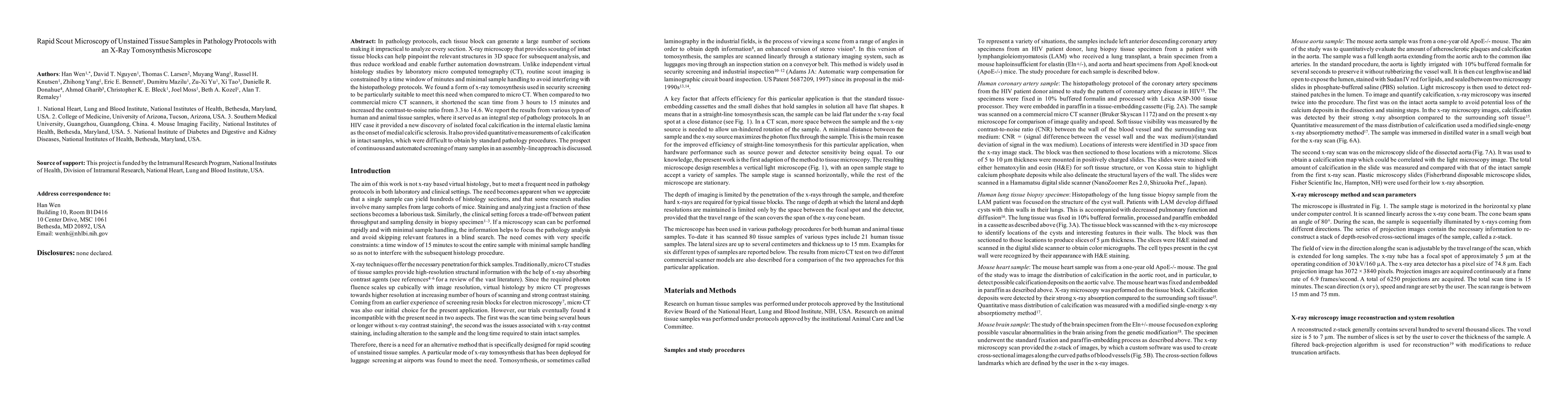
In pathology protocols, each tissue block can generate a large number of sections making it impractical to analyze every section. X-ray microscopy that provides a rapid survey of intact tissue blocks can help pinpoint the relevant structures in 3D space for subsequent analysis, and thus reduce workload and enable further automation downstream. Unlike dedicated virtual histology studies by traditional micro computed tomography (CT), routine scout imaging is constrained by a time window of minutes and minimal sample handling to avoid interfering with the pathology protocols. Traditional micro CT was not able to meet the requirements due to lengthy study times or sample alteration by the introduction of x-ray contrast agents. A form of x-ray tomosynthesis used in security screening was found to be efficient for rapid microscopy of unstained samples. When compared to a commercial micro CT scanner, it provided a 10-fold increase in imaging speed and a 4.8-fold increase in contrast-to-noise ratio. We report the results from a variety of human and animal tissue samples, where it served as an integral step of pathology protocols. In cases of vascular disease, it provided quantitative measurements of calcification in intact samples, which were difficult to obtain by standard pathology procedures. The prospect of continuous and automated screening of many samples in an assembly-line approach is discussed.
AI Key Findings
Get AI-generated insights about this paper's methodology, results, significance, and more — seven facets brought into focus.
Impact
Paper Details
Authors
PDF Preview
Key Terms
Citation Network
Current paper (gray), citations (green), references (blue)
Display is limited for performance on very large graphs.
Discussion 0