Summary
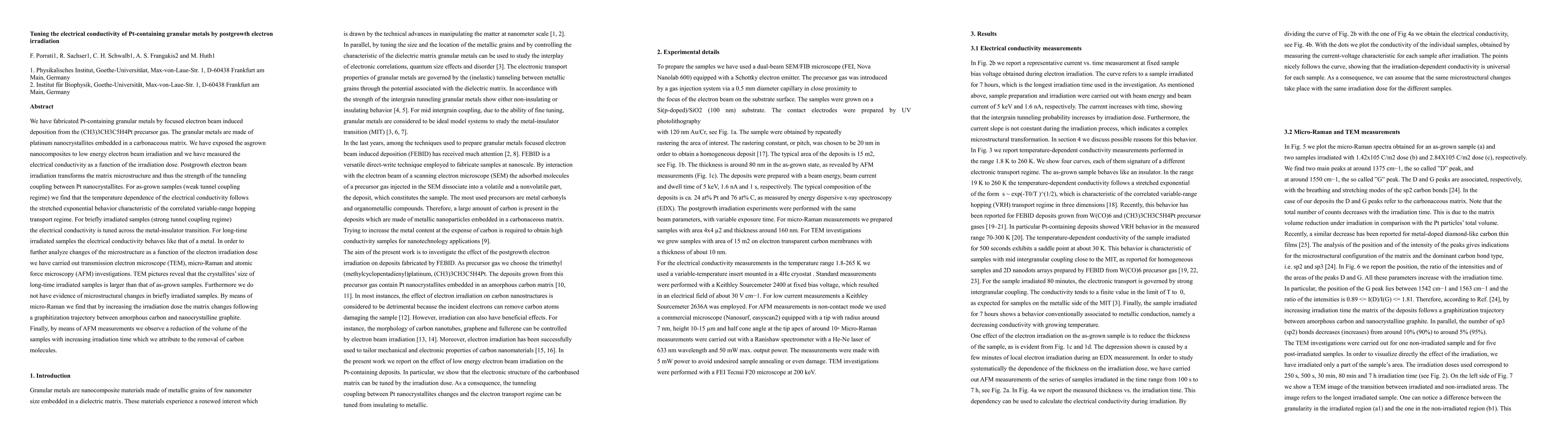
We have fabricated Pt-containing granular metals by focused electron beam induced deposition from the $(CH_3)_3CH_3C_5H_4Pt$ precursor gas. The granular metals are made of platinum nanocrystallites embedded in a carbonaceous matrix. We have exposed the as-grown nanocomposites to low energy electron beam irradiation and we have measured the electrical conductivity as a function of the irradiation dose. Postgrowth electron beam irradiation transforms the matrix microstructure and thus the strength of the tunneling coupling between Pt nanocrystallites. For as-grown samples (weak tunnel coupling regime) we find that the temperature dependence of the electrical conductivity follows the stretched exponential behavior characteristic of the correlated variable-range hopping transport regime. For briefly irradiated samples (strong tunnel coupling regime) the electrical conductivity is tuned across the metal-insulator transition. For long-time irradiated samples the electrical conductivity behaves like that of a metal. In order to further analyze changes of the microstructure as a function of the electron irradiation dose we have carried out transmission electron microscope (TEM), micro-Raman and atomic force microscopy (AFM) investigations. TEM pictures reveal that the crystallites' size of long-time irradiated samples is larger than that of as-grown samples. Furthermore we do not have evidence of microstructural changes in briefly irradiated samples. By means of micro-Raman we find that by increasing the irradiation dose the matrix changes following a graphitization trajectory between amorphous carbon and nanocrystalline graphite. Finally, by means of AFM measurements we observe a reduction of the volume of the samples with increasing irradiation time which we attribute to the removal of carbon molecules.
AI Key Findings
Get AI-generated insights about this paper's methodology, results, and significance.
Paper Details
PDF Preview
Key Terms
Citation Network
Current paper (gray), citations (green), references (blue)
Display is limited for performance on very large graphs.
| Title | Authors | Year | Actions |
|---|
Comments (0)