Two-Phase Segmentation Approach for Accurate Left Ventricle Segmentation in Cardiac MRI using Machine Learning
Publication
Metrics
AI Quick Summary
This research proposes a two-phase segmentation approach to enhance the accurate segmentation of the Left Ventricle (LV) in Cardiac MRI (CMR) scans using Machine Learning. The method addresses the challenge of parameter optimization by employing distinct parameters for Basal, Mid-Ventricle, and Apical LV slices, achieving a mean score of 0.9228 via 10-Fold Cross Validation on the ACDC dataset, demonstrating improved LV segmentation accuracy.
Paper Preview
Abstract
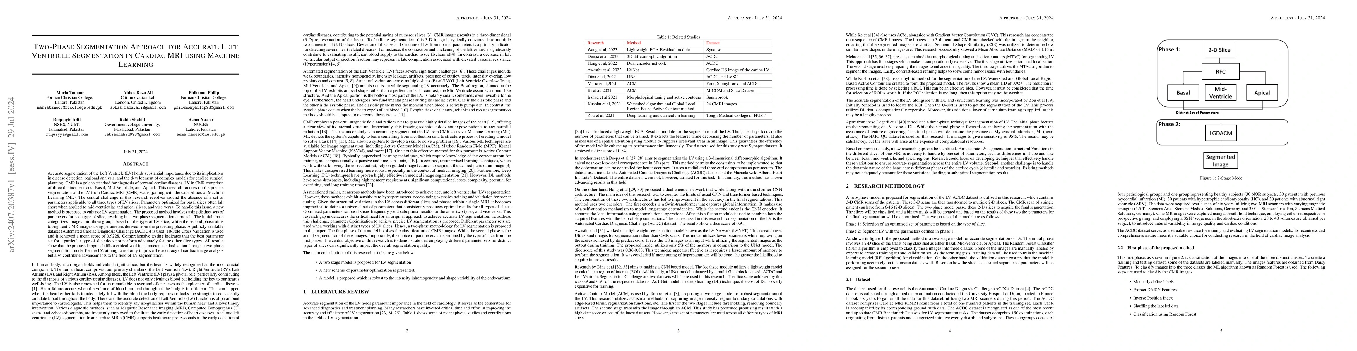
Accurate segmentation of the Left Ventricle (LV) holds substantial importance due to its implications in disease detection, regional analysis, and the development of complex models for cardiac surgical planning. CMR is a golden standard for diagnosis of serveral cardiac diseases. LV in CMR comprises of three distinct sections: Basal, Mid-Ventricle, and Apical. This research focuses on the precise segmentation of the LV from Cardiac MRI (CMR) scans, joining with the capabilities of Machine Learning (ML). The central challenge in this research revolves around the absence of a set of parameters applicable to all three types of LV slices. Parameters optimized for basal slices often fall short when applied to mid-ventricular and apical slices, and vice versa. To handle this issue, a new method is proposed to enhance LV segmentation. The proposed method involves using distinct sets of parameters for each type of slice, resulting in a two-phase segmentation approach. The initial phase categorizes images into three groups based on the type of LV slice, while the second phase aims to segment CMR images using parameters derived from the preceding phase. A publicly available dataset (Automated Cardiac Diagnosis Challenge (ACDC)) is used. 10-Fold Cross Validation is used and it achieved a mean score of 0.9228. Comprehensive testing indicates that the best parameter set for a particular type of slice does not perform adequately for the other slice types. All results show that the proposed approach fills a critical void in parameter standardization through a two-phase segmentation model for the LV, aiming to not only improve the accuracy of cardiac image analysis but also contribute advancements to the field of LV segmentation.
AI Key Findings
Get AI-generated insights about this paper's methodology, results, significance, and more — seven facets brought into focus.
Impact
Authors
PDF Preview
Key Terms
Citation Network
Current paper (gray), citations (green), references (blue)
Display is limited for performance on very large graphs.
Discussion 0