Summary
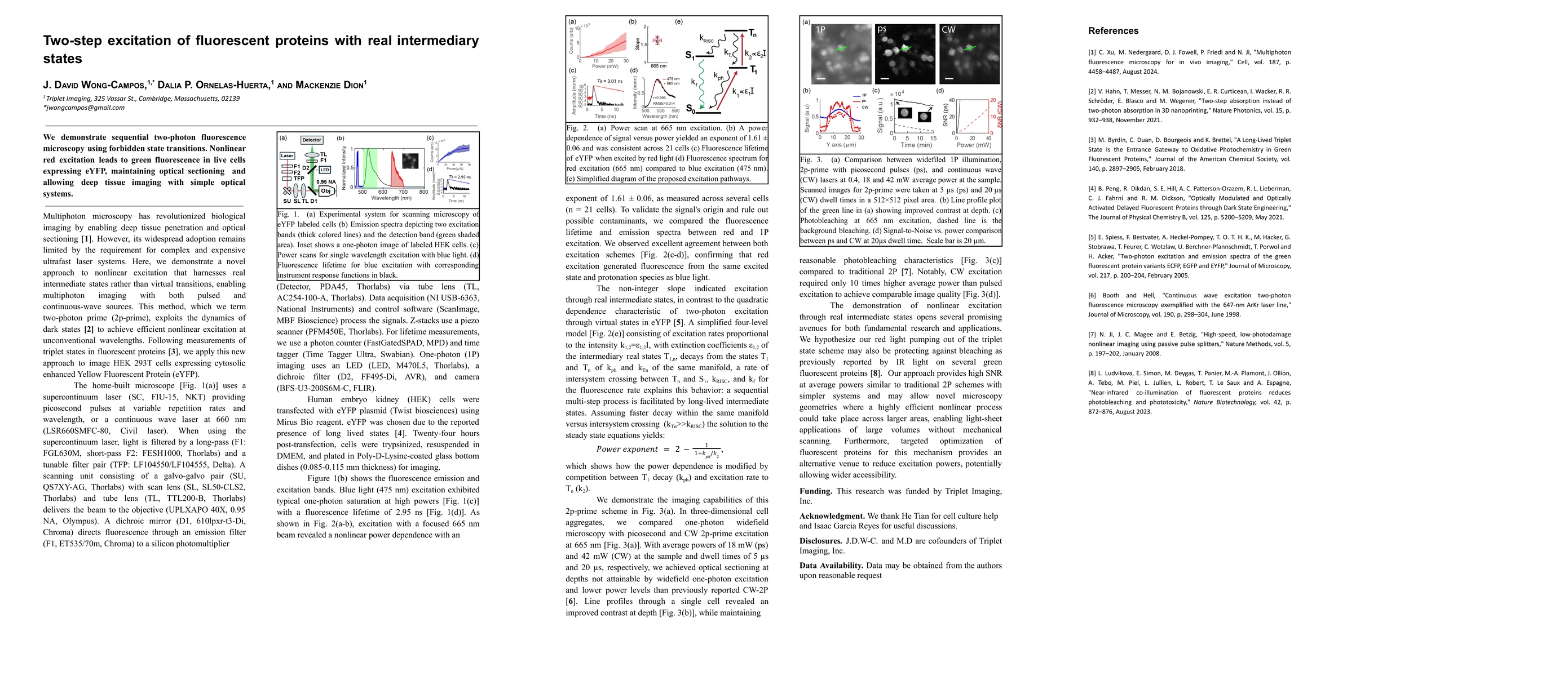
We demonstrate sequential two-photon fluorescence microscopy using forbidden state transitions. Nonlinear red excitation leads to green fluorescence in live cells expressing eYFP, maintaining optical sectioning and allowing deep tissue imaging with simple optical systems.
AI Key Findings
Generated Jun 10, 2025
Methodology
The research demonstrates sequential two-photon fluorescence microscopy using forbidden state transitions, exploiting real intermediate states for nonlinear excitation at unconventional wavelengths with both pulsed and continuous-wave sources.
Key Results
- Nonlinear red excitation leads to green fluorescence in live cells expressing eYFP.
- Optical sectioning and deep tissue imaging are maintained with simple optical systems.
- 2p-prime scheme achieves optical sectioning at greater depths than widefield one-photon excitation and lower power levels than previously reported CW-2P.
- CW excitation requires only 10 times higher average power than pulsed excitation to achieve comparable image quality.
Significance
This novel approach to nonlinear excitation harnesses real intermediate states, enabling multiphoton imaging with simpler and less expensive optical systems, which could revolutionize biological imaging by making it more accessible.
Technical Contribution
The introduction of the 'two-photon prime' (2p-prime) method, which utilizes real intermediate states for nonlinear excitation, enabling multiphoton imaging with both pulsed and continuous-wave sources.
Novelty
This work differs from existing research by employing real intermediate states instead of virtual transitions for multiphoton excitation, allowing for efficient nonlinear excitation at unconventional wavelengths with simpler optical systems.
Limitations
- The study was conducted using a home-built microscope, which may limit its immediate applicability in standard laboratory settings.
- Further research is needed to validate the method's compatibility with a broader range of fluorescent proteins and biological samples.
Future Work
- Explore the applicability of this method with various fluorescent proteins and biological samples.
- Investigate the potential of this approach for light-sheet microscopy applications.
Paper Details
PDF Preview
Similar Papers
Found 4 papersMultiphoton fluorescence excitation with real intermediary states
J. David Wong-Campos, Mackenzie Dion
A novel tool for labeling intermediary proteins between two non-interacting proteins
Li, H., Xie, L., Wu, G. et al.
No citations found for this paper.

Comments (0)