U-Net-based Models for Skin Lesion Segmentation: More Attention and Augmentation
Publication
Metrics
AI Quick Summary
This paper explores U-Net-based models for skin lesion segmentation, comparing ten models and augmentation strategies on the ISIC 2016 dataset. The U-Net-Resnet50 and R2U-Net models, combined with specific augmentation techniques, achieve the highest performance metrics, while attention modules like CBAM and AG blocks significantly enhance segmentation results with minimal computational cost.
Paper Preview
Abstract
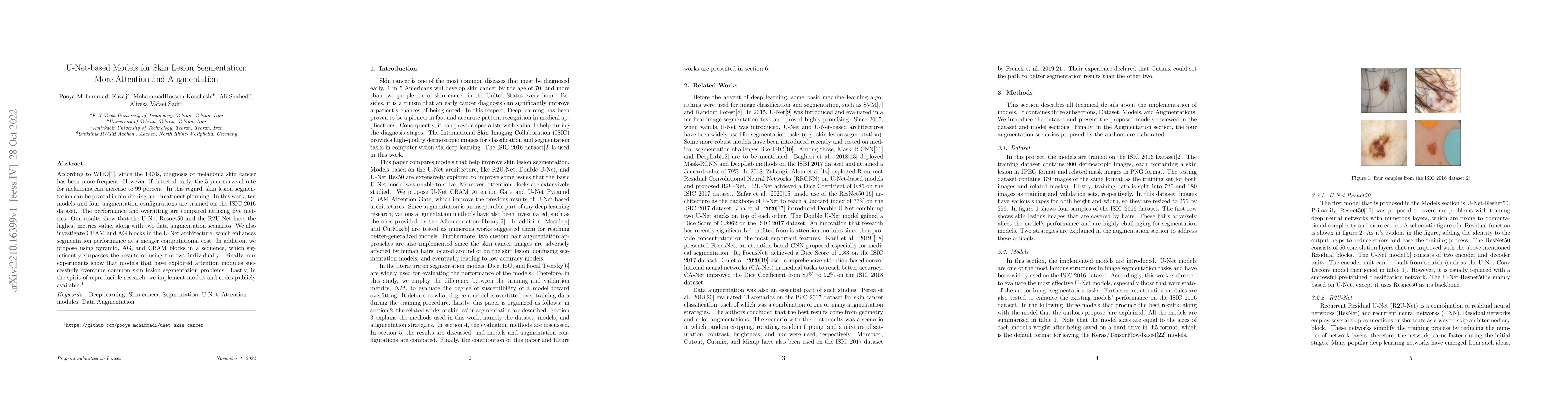
According to WHO[1], since the 1970s, diagnosis of melanoma skin cancer has been more frequent. However, if detected early, the 5-year survival rate for melanoma can increase to 99 percent. In this regard, skin lesion segmentation can be pivotal in monitoring and treatment planning. In this work, ten models and four augmentation configurations are trained on the ISIC 2016 dataset. The performance and overfitting are compared utilizing five metrics. Our results show that the U-Net-Resnet50 and the R2U-Net have the highest metrics value, along with two data augmentation scenarios. We also investigate CBAM and AG blocks in the U-Net architecture, which enhances segmentation performance at a meager computational cost. In addition, we propose using pyramid, AG, and CBAM blocks in a sequence, which significantly surpasses the results of using the two individually. Finally, our experiments show that models that have exploited attention modules successfully overcome common skin lesion segmentation problems. Lastly, in the spirit of reproducible research, we implement models and codes publicly available.
AI Key Findings
Get AI-generated insights about this paper's methodology, results, significance, and more — seven facets brought into focus.
Impact
Paper Details
Authors
PDF Preview
Key Terms
Citation Network
Current paper (gray), citations (green), references (blue)
Display is limited for performance on very large graphs.
Discussion 0