Publication

Published: Jan 06, 2026
Categories: cs.CV, cs.AI

Metrics

Source: ArXiv

Quick Actions

View PDF

Quick Answers

What methodology did the authors use?

The study introduces ULS+, an enhanced version of the Universal Lesion Segmentation (ULS) model, incorporating additional datasets and smaller input sizes. Training involved data-driven updates, click-point augmentation, and comparison against the original ULS model using Dice scores and robustness metrics. More in Methodology →

What are the key results?

ULS+ significantly outperformed the original ULS model in both Dice score and robustness across all tested datasets. — ULS+ achieved first place on the ULS23 Challenge test-phase leaderboard. More in Key Results →

Why is this work significant?

This research advances lesion segmentation in CT scans by improving accuracy and robustness, making the model more clinically relevant and applicable in real-world settings. The approach highlights the value of iterative model updates with new data for sustaining AI tool effectiveness. More in Significance →

What are the main limitations?

The smaller input size limits segmentation of lesions extending beyond the field of view. — Robustness scores were slightly lower for skeletal and undefined lesions due to structural challenges. More in Limitations →

Paper Preview

Abstract

In this study, we present ULS+, an enhanced version of the Universal Lesion Segmentation (ULS) model. The original ULS model segments lesions across the whole body in CT scans given volumes of interest (VOIs) centered around a click-point. Since its release, several new public datasets have become available that can further improve model performance. ULS+ incorporates these additional datasets and uses smaller input image sizes, resulting in higher accuracy and faster inference. We compared ULS and ULS+ using the Dice score and robustness to click-point location on the ULS23 Challenge test data and a subset of the Longitudinal-CT dataset. In all comparisons, ULS+ significantly outperformed ULS. Additionally, ULS+ ranks first on the ULS23 Challenge test-phase leaderboard. By maintaining a cycle of data-driven updates and clinical validation, ULS+ establishes a foundation for robust and clinically relevant lesion segmentation models.
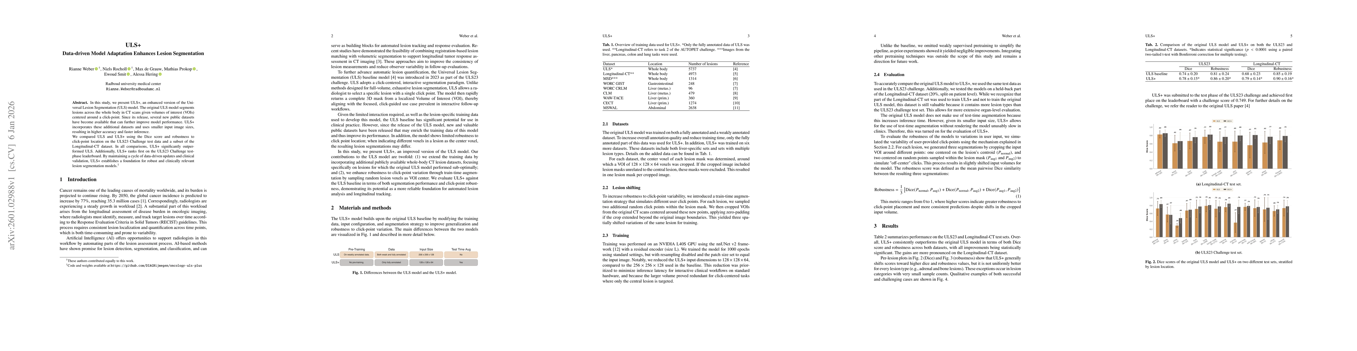
AI Key Findings

Generated Jan 07, 2026

Methodology — What approach did the authors take?

The study introduces ULS+, an enhanced version of the Universal Lesion Segmentation (ULS) model, incorporating additional datasets and smaller input sizes. Training involved data-driven updates, click-point augmentation, and comparison against the original ULS model using Dice scores and robustness metrics.

Key Results — What are the main findings?

  • ULS+ significantly outperformed the original ULS model in both Dice score and robustness across all tested datasets.
  • ULS+ achieved first place on the ULS23 Challenge test-phase leaderboard.
  • Performance improvements were more pronounced on the Longitudinal-CT dataset compared to ULS23.

Significance — Why does this research matter?

This research advances lesion segmentation in CT scans by improving accuracy and robustness, making the model more clinically relevant and applicable in real-world settings. The approach highlights the value of iterative model updates with new data for sustaining AI tool effectiveness.

Technical Contribution — What is the technical contribution?

ULS+ integrates expanded, fully annotated multi-organ data, click-point augmentation, and a reduced input size with test-time augmentation, enhancing both performance and speed.

Novelty — What is new about this work?

The work introduces a data-driven model adaptation framework that combines multiple datasets, click-point variability simulation, and smaller input sizes to achieve better segmentation accuracy and robustness compared to previous methods.

Limitations — What are the limitations of this study?

  • The smaller input size limits segmentation of lesions extending beyond the field of view.
  • Robustness scores were slightly lower for skeletal and undefined lesions due to structural challenges.

Future Work — What did the authors propose for future work?

  • Conducting a systematic ablation study to isolate the contribution of each improvement factor.
  • Exploring sliding-window inference strategies to address segmentation limitations with smaller input sizes.

Paper Details

Paper ID: 2601.02988
Comments: Accepted for publication at BVM 2026 (Bildverarbeitung für die Medizin), peer-reviewed conference paper
Categories:
cs.CV cs.AI

How to Cite This Paper

BibTeX
@article{hering2026uls,
  title         = {ULS+: Data-driven Model Adaptation Enhances Lesion Segmentation},
  author        = {Hering, Alessa and Smit, Ewoud and Prokop, Mathias and others},
  year          = {2026},
  eprint        = {2601.02988},
  archivePrefix = {arXiv},
  primaryClass  = {cs.CV},
}
APA 7
Hering, A., Smit, E., Prokop, M., Grauw, M., Rocholl, N., & Weber, R. (2026). ULS+: Data-driven Model Adaptation Enhances Lesion Segmentation. arXiv. https://arxiv.org/abs/2601.02988
MLA 9
Hering, Alessa, et al. "ULS+: Data-driven Model Adaptation Enhances Lesion Segmentation." arXiv, 2026, arxiv.org/abs/2601.02988.

Comments (0)

This website uses cookies to ensure you get the best experience. Learn more