This paper presents holiAtlas, a high-resolution multimodal brain atlas that integrates T1, T2, and WMn MRI images, achieving 0.125 mm³ resolution. It provides a dense labelling protocol for detailed brain anatomy from organ to substructure levels, constructed from 75 healthy subjects' data.
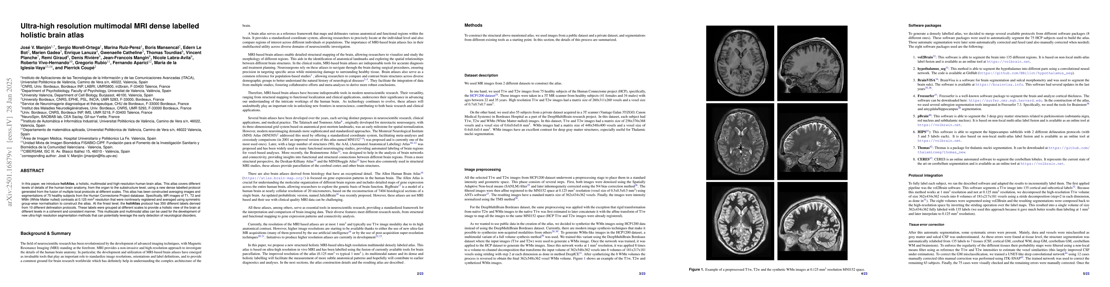
In this paper, we introduce holiAtlas, a holistic, multimodal and high-resolution human brain atlas. This atlas covers different levels of details of the human brain anatomy, from the organ to the substructure level, using a new dense labelled protocol generated from the fusion of multiple local protocols at different scales. This atlas has been constructed averaging images and segmentations of 75 healthy subjects from the Human Connectome Project database. Specifically, MR images of T1, T2 and WMn (White Matter nulled) contrasts at 0.125 $mm^{3}$ resolution that were nonlinearly registered and averaged using symmetric group-wise normalisation to construct the atlas. At the finest level, the holiAtlas protocol has 350 different labels derived from 10 different delineation protocols. These labels were grouped at different scales to provide a holistic view of the brain at different levels in a coherent and consistent manner. This multiscale and multimodal atlas can be used for the development of new ultra-high resolution segmentation methods that can potentially leverage the early detection of neurological disorders.
Get AI-generated insights about this paper's methodology, results, and significance.
Current paper (gray), citations (green), references (blue)
No similar papers found for this research.
Title | Authors | Year | Actions |
---|---|---|---|
DeepThalamus: A novel deep learning method for automatic segmentation of brain thalamic nuclei from multimodal ultra-high resolution MRI
|
Marina Ruiz-Perez, Sergio Morell-Ortega, Marien Gadea, Roberto Vivó Hernando, Gregorio Rubio, F. Aparici-Robles, M. Iglesia-Vayá, T. Tourdias, Pierrick Coup'e, J. V. Manj'on
|
2024
|
View Paper |
Title | Authors | Year | Actions |
---|---|---|---|
Staging of progressive supranuclear palsy-Richardson syndrome using MRI brain charts for the human lifespan
|
V. Planche, Boris Mansencal, J. Manjón, Wassilios G Meissner, T. Tourdias, P. Coupé
|
2024
|
View Paper |
DeepCERES: A deep learning method for cerebellar lobule segmentation using ultra-high resolution multimodal MRI
|
Sergio Morell-Ortega, Marina Ruiz-Perez, Marien Gadea, Roberto Vivó Hernando, Gregorio Rubio, F. Aparici-Robles, M. Iglesia-Vayá, G. Catheline, Pierrick Coup'e, J. V. Manj'on
|
2024
|
View Paper |
Emerging Trends in Fast MRI Using Deep-Learning Reconstruction on Undersampled k-Space Data: A Systematic Review
|
Dilbag Singh, Anmol Monga, H. D. de Moura, Xiaoxia Zhang, M. Zibetti, R. Regatte
|
2023
|
View Paper |
Human amygdala involvement in Alzheimer's disease revealed by stereological and dia‐PASEF analysis
|
Melania Gonzalez-Rodriguez, Sandra Villar-Conde, Veronica Astillero-Lopez, Patricia Villanueva-Anguita, I. Úbeda-Banon, A. Flores-Cuadrado, A. Martínez-Marcos, D. Saiz-Sánchez
|
2023
|
View Paper |
Lifespan neurodegeneration of the human brain in multiple sclerosis
|
P. Coupé, V. Planche, Boris Mansencal, Reda A. Kamroui, Ismail Koubiyr, J. Manjón, T. Tourdias
|
2023
|
View Paper |
Anatomical MRI staging of frontotemporal dementia variants
|
V. Planche, Boris Mansencal, J. Manjón, T. Tourdias, G. Catheline, P. Coupé
|
2023
|
View Paper |
HumanBrainAtlas: an in vivo MRI dataset for detailed segmentations
|
M. Schira, Zoey J. Isherwood, M. Kassem, M. Barth, T. Shaw, Michelle M Roberts, G. Paxinos
|
2022
|
View Paper |
Structural progression of Alzheimer’s disease over decades: the MRI staging scheme
|
V. Planche, J. Manjón, Boris Mansencal, E. Lanuza, T. Tourdias, G. Catheline, P. Coupé
|
2022
|
View Paper |
Browsing Multiple Subjects When the Atlas Adaptation Cannot Be Achieved via a Warping Strategy
|
D. Rivière, Yann Leprince, N. Labra, Nabil Vindas, O. Foubet, Bastien Cagna, K. Loh, Will Hopkins, A. Balzeau, M. Mancip, J. Lebenberg, Y. Cointepas, O. Coulon, J. F. Mangin
|
2022
|
View Paper |
vol2Brain: A New Online Pipeline for Whole Brain MRI Analysis
|
J. Manjón, José E. Romero, Roberto Vivó Hernando, G. Rubio, F. Aparici-Robles, M. Iglesia-Vayá, P. Coupé
|
2022
|
View Paper |
Automated segmentation of the hypothalamus and associated subunits in brain MRI
|
Benjamin Billot, M. Bocchetta, E. Todd, Adrian V. Dalca, J. E. Iglesias
|
2020
|
View Paper |
Julich-Brain: A 3D probabilistic atlas of the human brain’s cytoarchitecture
|
K. Amunts, H. Mohlberg, S. Bludau, K. Zilles
|
2020
|
View Paper |
Automated anatomical labelling atlas 3
|
E. Rolls, Chu-Chung Huang, Ching-Po Lin, Jianfeng Feng, M. Joliot
|
2020
|
View Paper |
Deep ICE: A Deep learning approach for MRI Intracranial Cavity Extraction.
|
J. V. Manj'on, José E. Romero, Roberto Vivo-Hernando, Gregorio Rubio-Navarro, M. Iglesia-Vayá, F. Aparici-Robles, Pierrick Coup'e
|
2020
|
View Paper |
pBrain: A novel pipeline for Parkinson related brain structure segmentation
|
J. Manjón, Alexa Berto, José E. Romero, E. Lanuza, Roberto Vivo-Hernando, F. Aparici-Robles, P. Coupé
|
2020
|
View Paper |
AssemblyNet: A large ensemble of CNNs for 3D whole brain MRI segmentation
|
P. Coupé, Boris Mansencal, Michaël Clément, Rémi Giraud, B. D. D. Senneville, T. Thong, V. Lepetit, J. Manjón
|
2019
|
View Paper |
Thalamus Optimized Multi Atlas Segmentation (THOMAS): fast, fully automated segmentation of thalamic nuclei from structural MRI
|
Jason H Su, Francis T. Thomas, W. Kasoff, T. Tourdias, Eun Young Choi, B. Rutt, M. Saranathan
|
2019
|
View Paper |
Lifespan Changes of the Human Brain In Alzheimer’s Disease
|
P. Coupé, J. Manjón, E. Lanuza, G. Catheline
|
2019
|
View Paper |
Image Synthesis in Multi-Contrast MRI With Conditional Generative Adversarial Networks
|
S. Dar, Mahmut Yurt, Levent Karacan, Aykut Erdem, Erkut Erdem, Tolga Cukur
|
2018
|
View Paper |
HIPS: A new hippocampus subfield segmentation method
|
José E. Romero, P. Coupé, J. Manjón
|
2017
|
View Paper |
High-resolution magnetic resonance imaging reveals nuclei of the human amygdala: manual segmentation to automatic atlas
|
Z. Saygin, Dorit Kliemann, J. E. Iglesias, A. Kouwe, Ellen Boyd, M. Reuter, A. Stevens, K. V. Leemput, Alexis McKee, M. Frosch, B. Fischl, J. Augustinack
|
2017
|
View Paper |
Striatal and Hippocampal Atrophy in Idiopathic Parkinson’s Disease Patients without Dementia: A Morphometric Analysis
|
Jared J. Tanner, Nikolaus R. McFarland, C. Price
|
2017
|
View Paper |
CERES: A new cerebellum lobule segmentation method
|
José E. Romero, P. Coupé, Rémi Giraud, Vinh-Thong Ta, Vladimir S Fonov, M. T. Park, M. Chakravarty, A. Voineskos, J. Manjón
|
2017
|
View Paper |
Mindboggling morphometry of human brains
|
A. Klein, Satrajit S. Ghosh, F. S. Bao, J. Giard, Yrjö Häme, E. Stavsky, Noah Lee, Brian Rossa, M. Reuter, E. C. Neto, A. Keshavan
|
2016
|
View Paper |
Implementation of a new parcellation of the orbitofrontal cortex in the automated anatomical labeling atlas
|
E. Rolls, M. Joliot, N. Tzourio-Mazoyer
|
2015
|
View Paper |
Bayesian segmentation of brainstem structures in MRI
|
J. E. Iglesias, K. V. Leemput, P. Bhatt, Christen Casillas, Shubir Dutt, N. Schuff, Diana Truran-Sacrey, A. Boxer, B. Fischl
|
2015
|
View Paper |
A computational atlas of the hippocampal formation using ex vivo, ultra-high resolution MRI: Application to adaptive segmentation of in vivo MRI
|
J. E. Iglesias, J. Augustinack, Khoa Nguyen, Christopher M. Player, Allison Player, M. Wright, N. Roy, M. Frosch, A. Mckee, L. Wald, B. Fischl, K. V. Leemput
|
2015
|
View Paper |
Large-scale evaluation of ANTs and FreeSurfer cortical thickness measurements
|
N. Tustison, P. Cook, A. Klein, G. Song, Sandhitsu R. Das, J. Duda, Benjamin M. Kandel, N. V. Strien, J. Stone, J. Gee, B. Avants
|
2014
|
View Paper |
A novel in vivo atlas of human hippocampal subfields using high-resolution 3T magnetic resonance imaging
|
Julie L. Winterburn, J. Pruessner, S. Chavez, M. Schira, N. Lobaugh, A. Voineskos, M. Chakravarty
|
2013
|
View Paper |
BigBrain: An Ultrahigh-Resolution 3D Human Brain Model
|
K. Amunts, C. Lepage, L. Borgeat, H. Mohlberg, Timo Dickscheid, Marc-Etienne Rousseau, S. Bludau, P. Bazin, L. Lewis, A. Oros-Peusquens, N. Shah, T. Lippert, K. Zilles, Alan C. Evans
|
2013
|
View Paper |
An anatomically comprehensive atlas of the adult human brain transcriptome
|
M. Hawrylycz, E. Lein, A. Guillozet‐Bongaarts, E. Shen, Lydia Ng, Jeremy A. Miller, L. N. V. D. Lagemaat, Kimberly A. Smith, Amanda J. Ebbert, Z. Riley, Chris Abajian, C. Beckmann, Amy Bernard, Darren Bertagnolli, A. Boe, P. Cartagena, M. Chakravarty, Mike Chapin, Jimmy Chong, R. Dalley, B. Daly, Chinh Dang, S. Datta, N. Dee, Tim Dolbeare, V. Faber, David Feng, David R. Fowler, J. Goldy, B. Gregor, Zeb Haradon, D. Haynor, J. Hohmann, S. Horvath, Robert E. Howard, A. Jeromin, Jayson M. Jochim, M. Kinnunen, C. Lau, E. Lazarz, Changkyu Lee, Tracy A. Lemon, Lingling Li, Yang Li, J. Morris, C. Overly, Patrick D. Parker, Sheana E. Parry, M. Reding, J. Royall, J. Schulkin, P. A. Sequeira, C. Slaughterbeck, Simon C. Smith, Andrew Sodt, S. Sunkin, B. Swanson, M. Vawter, Derric Williams, Paul E. Wohnoutka, H. Zielke, D. Geschwind, P. Hof, Stephen M. Smith, Christof Koch, S. Grant, Allan R. Jones
|
2012
|
View Paper |
Cortical sulci recognition and spatial normalization
|
M. Perrot, D. Rivière, J. F. Mangin
|
2011
|
View Paper |
Unbiased average age-appropriate atlases for pediatric studies
|
Vladimir S Fonov, Alan C. Evans, K. Botteron, C. Almli, R. McKinstry, D. Collins
|
2011
|
View Paper |
N4ITK: Improved N3 Bias Correction
|
N. Tustison, B. Avants, P. Cook, Yuanjie Zheng, Alexander Egan, Paul Yushkevich, J. Gee
|
2010
|
View Paper |
Adaptive non‐local means denoising of MR images with spatially varying noise levels
|
J. Manjón, P. Coupé, Luis Martí-Bonmatí, D. L. Collins, Montserrat Robles
|
2010
|
View Paper |
Unbiased nonlinear average age-appropriate brain templates from birth to adulthood
|
V. Fonov, A. Evans, RC McKinstry, C. Almli, D. Collins
|
2009
|
View Paper |
Et al
|
P. Cochat, L. Vaucoret, J. Sarles
|
2008
|
View Paper |
Robust MRI brain tissue parameter estimation by multistage outlier rejection
|
J. Manjón, Jussi Tohka, G. García-Martí, J. Carbonell-Caballero, J. J. Lull, L. Martí-Bonmatí, M. Robles
|
2008
|
View Paper |
Symmetric diffeomorphic image registration with cross-correlation: Evaluating automated labeling of elderly and neurodegenerative brain
|
B. Avants, C. Epstein, M. Grossman, J. Gee
|
2008
|
View Paper |
User-guided 3D active contour segmentation of anatomical structures: Significantly improved efficiency and reliability
|
Paul Yushkevich, J. Piven, H. Hazlett, R. G. Smith, Sean Ho, J. Gee, G. Gerig
|
2006
|
View Paper |
An automated labeling system for subdividing the human cerebral cortex on MRI scans into gyral based regions of interest
|
R. Desikan, F. Ségonne, B. Fischl, B. Quinn, B. Dickerson, D. Blacker, R. Buckner, A. Dale, R. P. Maguire, B. Hyman, M. Albert, R. Killiany
|
2006
|
View Paper |
Relationship between hippocampal volume and CA1 neuron loss in brains of humans with and without Alzheimer's disease
|
J. Kril, J. Hodges, G. Halliday
|
2004
|
View Paper |
A framework to study the cortical folding patterns
|
J. Mangin, D. Rivière, A. Cachia, E. Duchesnay, Y. Cointepas, D. Papadopoulos-Orfanos, P. Scifo, T. Ochiai, F. Brunelle, J. Régis
|
2004
|
View Paper |
Automated Anatomical Labeling of Activations in SPM Using a Macroscopic Anatomical Parcellation of the MNI MRI Single-Subject Brain
|
N. Tzourio-Mazoyer, B. Landeau, D. Papathanassiou, F. Crivello, O. Etard, N. Delcroix, B. Mazoyer, M. Joliot
|
2002
|
View Paper |
Measuring the thickness of the human cerebral cortex from magnetic resonance images.
|
B. Fischl, A. M. Dale
|
2000
|
View Paper |
The CA1 Region of the Human Hippocampus Is a Hot Spot in Alzheimer's Disease
|
M. West, C. Kawas, L. Martin, J. Troncoso
|
2000
|
View Paper |
3D statistical neuroanatomical models from 305 MRI volumes
|
Alan C. Evans, D. Collins, S. R. Mills, E. D. Brown, R. L. Kelly, T. Peters
|
1993
|
View Paper |
Co-Planar Stereotaxic Atlas of the Human Brain: 3-Dimensional Proportional System: An Approach to Cerebral Imaging
|
J. Talairach, P. Tournoux, Mark Rayport
|
1988
|
View Paper |
This method is able to segment the hypothalamus into different parts using a convolutional neural network
|
2020
|
||
This software is able to segment the hippocampus subfields with 2 different delineation protocols (with 3 and 5 labels each). It is also based on non-local multi-atlas label fusion and
|
2017
|
||
A Probabilistic Atlas and Reference System for the Human Brain
|
J. Mazziotta
|
2001
|
View Paper |
Freesurfer is a well-known software package to segment the brain and analyze cortical thickness
|
2000
|
||
2024. Super-resolution neural networks improve the spatiotemporal resolution of adaptive MRI-guided radiation therapy
|
|||
V. 2024. DeepThalamus: A novel deep learning method for automatic segmentation of brain thalamic nuclei from multimodal ultra-high resolution MRI
|
|||
2023. Super-resolution of brain MRI images based on denoising diffusion probabilistic model
|
|||
2024. A next-generation, histological atlas of the human brain and its application to automated brain MRI segmentation
|
Showing first page preview. Click "Open Full PDF" to view the complete paper.
Your comment contains inappropriate language and has been rejected.
Are you sure you want to remove this tag? This action cannot be undone.
Someone just favorited a paper!
Comments (0)