Summary
Intracortical US imaging extends B-mode imaging into bone using a dedicated image reconstruction algorithm that corrects for refraction at the bone-soft tissue interfaces. It has shown promising results in a few healthy, predominantly young adults, providing anatomical images of the cortex (periosteal and endosteal surfaces) along with estimations of US wave speed. However, its reliability in older or osteoporotic bones remains uncertain. In this study, we critically assessed the performance of intracortical US imaging ex vivo in bones with various microstructural patterns, including bones exhibiting signs of unbalanced intracortical remodeling. We analyzed factors influencing US image quality, particularly endosteal surface reconstruction, as well as the accuracy of wave speed estimation and its relationship with porosity. We imaged 20 regions of interest from the femoral diaphysis of five elderly donors using a 2.5 MHz US transducer. The reconstructed US images were compared to site-matched high-resolution micro-CT (HR-muCT) images. In samples with moderate porosity, the endosteal surface was accurately identified, and thickness estimates from US and HR-muCT differed by less than 10%. In highly remodeled bones with increased porosity, the reconstructed endosteal surface appeared less bright and was located above the cortex region containing resorption cavities. We observed a decrease in US wave speed with increasing cortical porosity suggesting that the method could discriminate between bones with low porosity (less than 5%) and those with moderate to high porosity (greater than ~10%). This study paves the way for the application of US imaging in diagnosing cortical bone health, particularly for detecting increased cortical porosity and reduced cortical thickness.
AI Key Findings
Generated Jun 11, 2025
Methodology
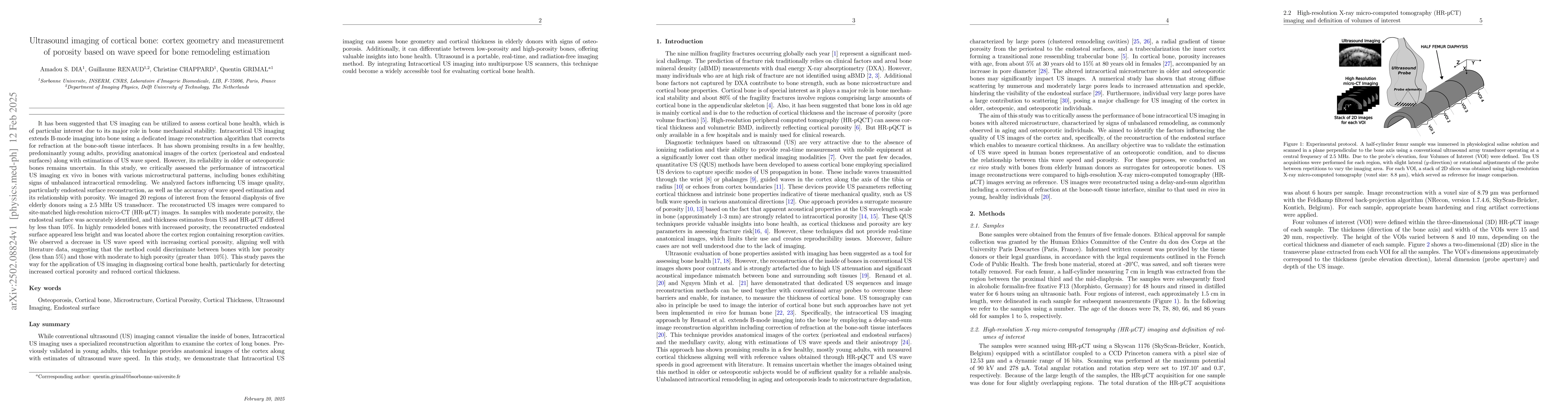
The study employs a critical evaluation of ultrasound (US) imaging for cortical bone assessment in elderly individuals, utilizing a 2.5 MHz phased-array probe and a delay-and-sum reconstruction algorithm to correct for refraction at bone-soft tissue interfaces. High-resolution micro-CT (HR-muCT) images serve as a reference for comparison.
Key Results
- US imaging accurately reconstructs cortical boundaries and measures cortical thickness in homogeneous and moderately porous samples.
- In highly porous samples, the endosteal surface brightness decreases, and its delineation may be ambiguous due to large pores, potentially indicating advanced cortical bone alteration.
- Wavespeed within cortical bone decreases with increasing porosity, suggesting the method can discriminate between bones with low porosity (<5%) and those with moderate to high porosity (>~10%).
- US imaging combined with the autofocus method provides reliable estimation of wavespeed in cortical bone samples with moderate microstructural heterogeneity.
- The study demonstrates the potential of US imaging to assess bone health in elderly or osteoporotic individuals by evaluating cortical thickness and cortical bone tissue mechanical competence, reflected by wave speed.
Significance
This research is significant as it validates the US imaging technique for diagnosing bone health, particularly for detecting increased cortical porosity and reduced cortical thickness, which are crucial factors in bone fracture risk.
Technical Contribution
The study presents a validated US imaging approach with Dijkstra segmentation for accurately reconstructing the endosteal boundary in elderly donors' bones, even with marked curvature, except in cases with very large pores.
Novelty
This work is novel as it critically assesses the potential of US imaging to diagnose bone health in elderly individuals, providing precise comparisons of geometric reconstructions against HR-muCT references and a critical evaluation of measured wave speed as it reflects porosity.
Limitations
- The study used femur bones due to availability constraints, which have a more pronounced curvature compared to other common US measurement sites like the tibia or radius.
- Only five bones were analyzed, with one being too porous for effective US imaging, highlighting the method's limitations in cases with large pores.
- The osteoporotic status of donors was unknown, although samples were classified into groups representing healthy, osteoporotic, and very advanced osteoporotic conditions.
Future Work
- Investigate the precision and sensitivity of US imaging in vivo in patients to determine its clinical applicability.
- Explore the implementation of this US imaging approach as an additional imaging mode on standard B-mode US systems, increasing its availability.
Paper Details
PDF Preview
Similar Papers
Found 4 papersRefraction corrected specular beamforming applied to cortical bone enhances interface visibility of bone-soft tissues interfaces
Quentin Grimal, Guillaume Renaud, Amadou S. Dia
No citations found for this paper.
Comments (0)