Ultrasound matrix imaging for transcranial in-vivo localization microscopy
Publication
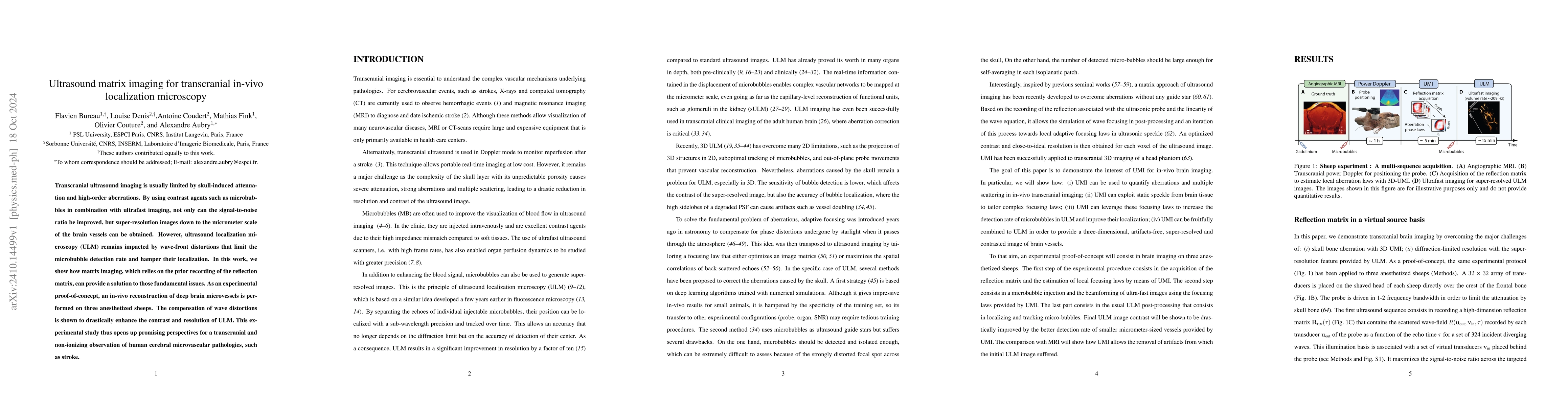
Metrics
AI Quick Summary
This paper explores the use of matrix imaging to enhance transcranial ultrasound localization microscopy (ULM) by compensating for wave-front distortions caused by the skull, significantly improving the resolution and contrast of brain microvascular imaging in anesthetized sheep, with potential applications for observing human cerebral pathologies like stroke.
Paper Preview
Abstract
Transcranial ultrasound imaging is usually limited by skull-induced attenuation and high-order aberrations. By using contrast agents such as microbubbles in combination with ultrafast imaging, not only can the signal-to-noise ratio be improved, but super-resolution images down to the micrometer scale of the brain vessels can be obtained. However, ultrasound localization microscopy (ULM) remains impacted by wave-front distortions that limit the microbubble detection rate and hamper their localization. In this work, we show how matrix imaging, which relies on the prior recording of the reflection matrix, can provide a solution to those fundamental issues. As an experimental proof-of-concept, an in-vivo reconstruction of deep brain microvessels is performed on three anesthetized sheeps. The compensation of wave distortions is shown to drastically enhance the contrast and resolution of ULM. This experimental study thus opens up promising perspectives for a transcranial and non-ionizing observation of human cerebral microvascular pathologies, such as stroke.
AI Key Findings
Get AI-generated insights about this paper's methodology, results, significance, and more — seven facets brought into focus.
Impact
Paper Details
Authors
PDF Preview
Citation Network
Current paper (gray), citations (green), references (blue)
Display is limited for performance on very large graphs.
Discussion 0