Publication
Metrics
AI Quick Summary
This paper presents a novel approach to disentangle the longitudinal relaxation times ($T_1$) of free and semi-solid spin pools in magnetization transfer imaging, finding that $T_1^s \ll T_1^f$ in healthy white matter. The results suggest MT significantly influences longitudinal relaxation and reveal larger semi-solid spin pool fractions, with significant differences in $T_1^f$ between relapsing-remitting MS patients and healthy controls.
Paper Preview
Abstract
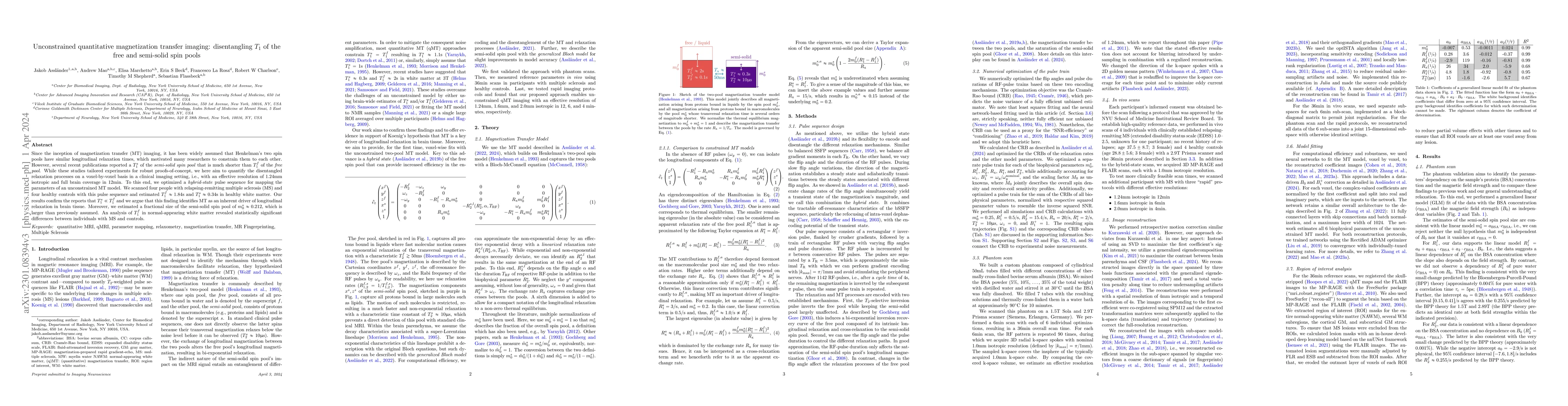
Since the inception of magnetization transfer (MT) imaging, it has been widely assumed that Henkelman's two spin pools have similar longitudinal relaxation times, which motivated many researchers to constrain them to each other. However, several recent publications reported a $T_1^s$ of the semi-solid spin pool that is much shorter than $T_1^f$ of the free pool. While these studies tailored experiments for robust proofs-of-concept, we here aim to quantify the disentangled relaxation processes on a voxel-by-voxel basis in a clinical imaging setting, i.e., with an effective resolution of 1.24mm isotropic and full brain coverage in 12min. To this end, we optimized a hybrid-state pulse sequence for mapping the parameters of an unconstrained MT model. We scanned four people with relapsing-remitting multiple sclerosis (MS) and four healthy controls with this pulse sequence and estimated $T_1^f \approx 1.84$s and $T_1^s \approx 0.34$s in healthy white matter. Our results confirm the reports that $T_1^s \ll T_1^f$ and we argue that this finding identifies MT as an inherent driver of longitudinal relaxation in brain tissue. Moreover, we estimated a fractional size of the semi-solid spin pool of $m_0^s \approx 0.212$, which is larger than previously assumed. An analysis of $T_1^f$ in normal-appearing white matter revealed statistically significant differences between individuals with MS and controls.
AI Key Findings
Get AI-generated insights about this paper's methodology, results, significance, and more — seven facets brought into focus.
Impact
Paper Details
Authors
PDF Preview
Key Terms
Citation Network
Current paper (gray), citations (green), references (blue)
Display is limited for performance on very large graphs.
Discussion 0