Detailed Breakdown
The Problem
In the world of medical AI, tools are typically hyper-specialized and disconnected. You have one type of AI model that excels at understanding—it can analyze a chest X-ray to detect pneumonia or generate a text report describing its findings. You have another, completely separate type of model that excels at generation—it can create a synthetic medical image from a text description ("show me an MRI of a healthy brain") or enhance a low-resolution scan.
This separation is a major bottleneck. A human radiologist's workflow is unified: they observe an image, consult their knowledge, and can both analyze it (write a report) and mentally visualize or even sketch out variations ("what if this shadow wasn't there?"). Existing AI systems break this fluid process into isolated tasks. This fragmentation means:
Inefficient Workflows: Hospitals and tech companies need to stitch together multiple, distinct AI systems to cover a full diagnostic pipeline.
Lost Synergy: The models can't learn from each other. The insights gained from generating realistic medical images could make an AI better at understanding real ones, but this connection is lost.
Limited Capabilities: A doctor can't ask an "understanding" model to visually highlight a region of concern or generate a "what-if" image to plan a surgery.
This research addresses the fundamental problem of creating a single, unified AI that can simultaneously understand and generate multimodal medical data, mirroring the comprehensive workflow of a human expert.
The Innovation
The key breakthrough of this paper is UniMedVL, the first medical AI model designed from the ground up to be truly unified. It doesn't just bolt an "understanding" module onto a "generation" module; it integrates both capabilities into a single, cohesive architecture.
What makes it different is its core philosophy, the Observation-Knowledge-Analysis (OKA) paradigm, which mimics a physician's diagnostic process:
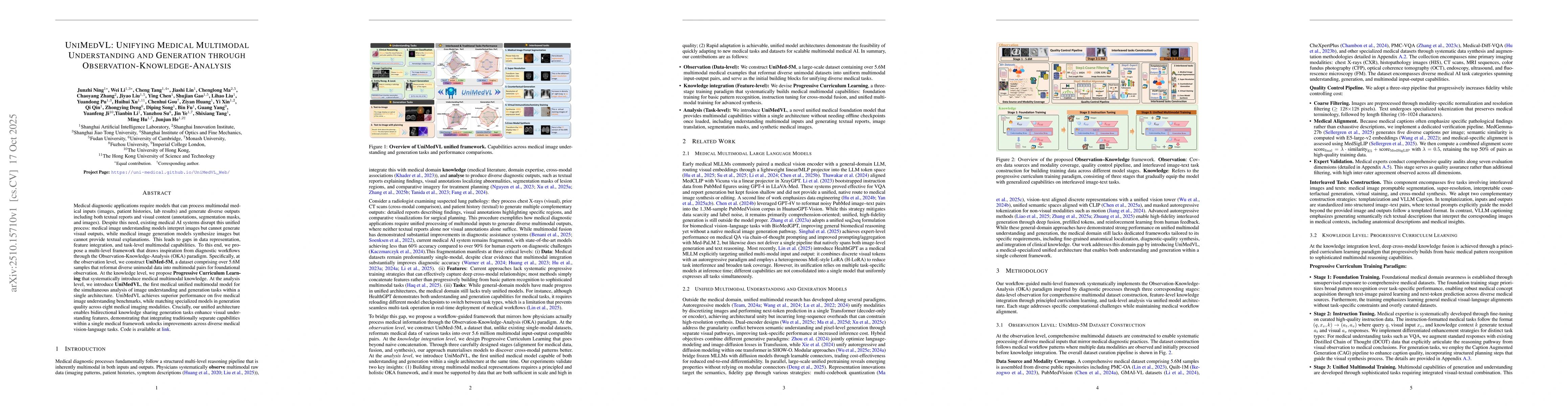
Observation (The Data): The authors first built UniMed-5M, a massive, curated dataset of over 5.6 million medical image-text samples. Crucially, they reformatted data from dozens of sources into a consistent format, creating a strong foundation for a unified model.
Knowledge (The Training): Instead of just throwing all the data at the model, they use Progressive Curriculum Learning. This is a staged training process that starts with basic pattern recognition (like a first-year med student) and gradually moves to complex, instruction-based tasks that combine understanding and generation (like an experienced specialist).
Analysis (The Model): The UniMedVL architecture itself is novel. It uses a "Mixture-of-Experts" approach, where different parts of the neural network specialize in either understanding or generation but operate within the same system. This allows the model to handle a vast range of tasks without needing to be reconfigured.
The most significant innovation is demonstrating bidirectional knowledge sharing: training the model on generation tasks actively improves its performance on understanding tasks, and vice-versa. This synergy was previously just a theory, but this paper provides strong evidence that it works in practice.
How It Works
UniMedVL is built using the three-stage OKA framework:
Step 1: Building the Foundation (Observation) The process starts with data. The team aggregated over 30 public datasets covering nine types of medical imaging (X-ray, CT, MRI, etc.). They then ran this data through a rigorous quality control pipeline, using other AI models to filter, align, and enrich the image-text pairs. This resulted in the UniMed-5M dataset, a high-quality, multimodal library ready for training.
Step 2: Smart, Staged Learning (Knowledge) The model learns progressively, like a human.
Foundation Training: The model is first exposed to the entire dataset to learn fundamental associations between medical images and text (e.g., what the word "fracture" looks like in an X-ray).
Instruction Tuning: Next, it's fine-tuned on specific, high-quality examples where it has to follow instructions, like "generate a report for this image" or "answer this question about the scan."
Unified Multimodal Training: Finally, it's trained on complex "interleaved" tasks that require both understanding and generation simultaneously. For example, given an image and the text "show this with increased pneumonia," the model must understand the input and generate a new, modified image.
Step 3: The Unified Architecture (Analysis) The UniMedVL model is a sophisticated Transformer-based system. Conceptually, it has two parallel pathways for processing images:
An Understanding Encoder: This part turns an image into a compact representation suitable for tasks like question answering and report generation.
A Generation Encoder/Decoder: This part processes an image into a detailed latent representation that can be used to reconstruct it or create a new image from scratch.
These pathways feed into a shared "Mixture-of-Experts" module. When a task comes in, the model intelligently routes the information to the relevant "expert" network—the understanding expert or the generation expert—all within one seamless architecture.
Key Results
UniMedVL demonstrated exceptional performance, proving a generalist model can compete with and even exceed specialists.
Superior Understanding: On five different medical image understanding benchmarks (like visual question answering), UniMedVL achieved state-of-the-art results among unified models. For instance, on the SLAKE benchmark, it scored 75.4% accuracy, outperforming the next-best unified model by a significant margin and approaching the performance of models built only for understanding.
Competitive Generation: In generating realistic medical images across eight modalities, UniMedVL's quality was on par with models designed exclusively for image generation.
Proven Synergy: The most crucial finding was that the full UniMedVL model, trained on both understanding and generation, produced significantly better images than a version of the model trained only on generation tasks. This confirms that the "understanding" knowledge directly enhances its "generation" capabilities.
Practical Applications
Next-Generation Radiologist Co-pilot: A tool that not only drafts a diagnostic report from a scan (understanding) but also allows the radiologist to ask for visual modifications. For example: "Generate a version of this CT scan where the suspected lesion is removed to help plan the surgery."
Synthetic Data Generation for Training: Medical schools and AI companies can use UniMedVL to create an almost infinite supply of realistic, diverse, and anonymized training data. A prompt like "Generate 100 chest X-rays showing varying stages of tuberculosis" can create a robust dataset without privacy concerns.
Cross-Modality Image Synthesis: In situations where a patient has an MRI but a CT scan is needed for analysis, this model could generate a high-fidelity synthetic CT from the MRI data. This could reduce costs, save time, and spare patients from additional radiation exposure.
Predictive Medical Visualization: For clinical trials, the model could take a baseline scan and a patient's data to generate a "counterfactual" image predicting the likely progression of a disease with or without a new treatment, offering a powerful visualization tool for researchers.
Limitations & Considerations
Focus on 2D Imaging: The current model primarily works with 2D images. Extending it to handle 3D volumetric data (like full 3D MRIs) and videos is a significant next step.
Clinical Validation: While the model performs exceptionally well on academic benchmarks, it has not undergone the rigorous, large-scale clinical trials required for real-world diagnostic use and regulatory approval (e.g., by the FDA).
Potential for Bias: The model's performance is dependent on the UniMed-5M dataset. Any inherent biases in that data regarding patient demographics, equipment, or disease prevalence could be learned and amplified by the model.
What This Means for Builders
For entrepreneurs and developers, UniMedVL signals a major paradigm shift in medical AI.
Move Beyond Single-Task Models: The future isn't about building the best "X-ray classifier" but about building comprehensive, multimodal foundation models. This research provides a blueprint and open-source code to start.
Embrace Data Synergy: When developing an AI product, consider how seemingly separate tasks can reinforce each other. If you're building a diagnostic tool, could adding a generative feature actually make your core diagnostic algorithm more robust? This paper says yes.
Leverage Open-Source Power: The authors have made their code available on GitHub. This is a massive accelerator. Instead of starting from scratch, builders can fine-tune UniMedVL on their proprietary data to create specialized commercial applications for radiology, pathology, ophthalmology, and more. The era of unified, multi-talented medical AI is here, and it's now accessible to build upon.
Discussion 0