Quick Answers
What methodology did the authors use?
Dual‑modal semantic alignment framework that injects class‑level structure into pixel‑wise learning via prototype and text branches operating in a shared embedding space, fused with visual decoder logits for pseudo‑label refinement. More in Methodology →
What are the key results?
Achieves Dice gains of +2.61% on GlaS and +8.64% on CRAG at 10% labeled data, with corresponding Jaccard improvements of +4.08% and +11.71%. — Prototype and text branches individually improve performance (+0.34% Dice, +0.53% Jaccard for prototype; +0.40% Dice, +0.62% Jaccard for text) and together yield the best results (+0.48% Dice, +0.76% Jaccard). More in Key Results →
Why is this work significant?
Provides a scalable, foundation‑model‑based approach that substantially improves semi‑supervised histopathology segmentation under limited annotations, enabling more accurate gland delineation and potentially reducing expert labeling effort. More in Significance →
What are the main limitations?
Relies on a frozen text encoder and fixed fusion weights, which may limit adaptability to other modalities or datasets. — Evaluation is confined to gland segmentation in colorectal histology; generalization to other organs or imaging modalities remains untested. More in Limitations →
Abstract
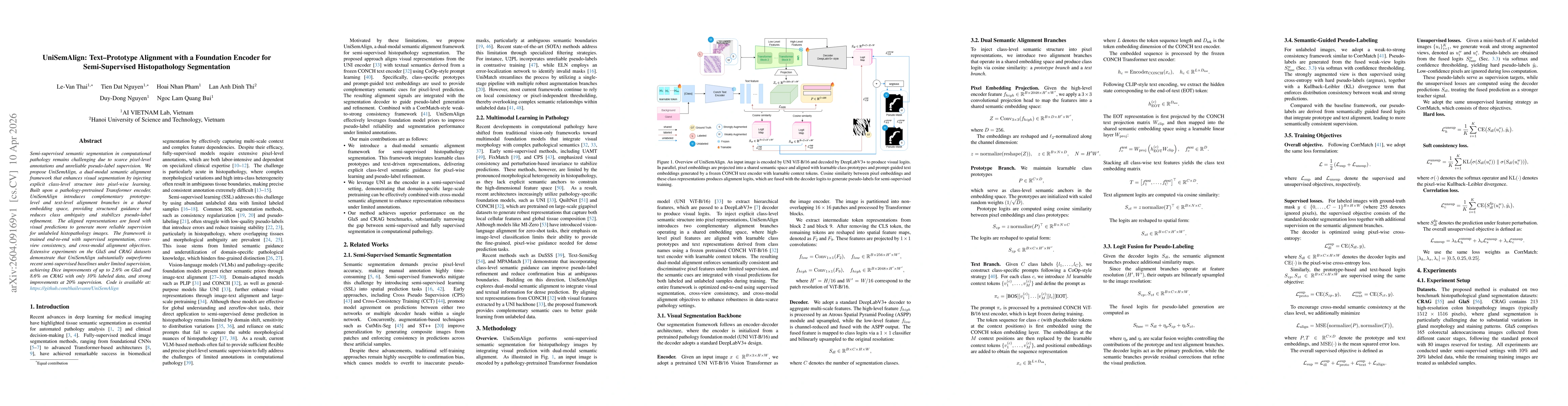
Semi-supervised semantic segmentation in computational pathology remains challenging due to scarce pixel-level annotations and unreliable pseudo-label supervision. We propose UniSemAlign, a dual-modal semantic alignment framework that enhances visual segmentation by injecting explicit class-level structure into pixel-wise learning. Built upon a pathology-pretrained Transformer encoder, UniSemAlign introduces complementary prototype-level and text-level alignment branches in a shared embedding space, providing structured guidance that reduces class ambiguity and stabilizes pseudo-label refinement. The aligned representations are fused with visual predictions to generate more reliable supervision for unlabeled histopathology images. The framework is trained end-to-end with supervised segmentation, cross-view consistency, and cross-modal alignment objectives. Extensive experiments on the GlaS and CRAG datasets demonstrate that UniSemAlign substantially outperforms recent semi-supervised baselines under limited supervision, achieving Dice improvements of up to 2.6% on GlaS and 8.6% on CRAG with only 10% labeled data, and strong improvements at 20% supervision. Code is available at: https://github.com/thailevann/UniSemAlign
AI Key Findings
Generated Apr 13, 2026
Methodology — What approach did the authors take?
Dual‑modal semantic alignment framework that injects class‑level structure into pixel‑wise learning via prototype and text branches operating in a shared embedding space, fused with visual decoder logits for pseudo‑label refinement.
Key Results — What are the main findings?
- Achieves Dice gains of +2.61% on GlaS and +8.64% on CRAG at 10% labeled data, with corresponding Jaccard improvements of +4.08% and +11.71%.
- Prototype and text branches individually improve performance (+0.34% Dice, +0.53% Jaccard for prototype; +0.40% Dice, +0.62% Jaccard for text) and together yield the best results (+0.48% Dice, +0.76% Jaccard).
- Ablation shows pathology‑pretrained UNI encoder outperforms ResNet101 by +1.77% Dice and +2.79% Jaccard, and MSE alignment loss outperforms cosine and KL losses by +0.58% Dice and +0.93% Jaccard.
Significance — Why does this research matter?
Provides a scalable, foundation‑model‑based approach that substantially improves semi‑supervised histopathology segmentation under limited annotations, enabling more accurate gland delineation and potentially reducing expert labeling effort.
Technical Contribution — What is the technical contribution?
Introduces a dual‑modal semantic alignment mechanism that fuses prototype‑based and text‑driven embeddings with visual predictions, enabling structured pseudo‑label generation and improved consistency in semi‑supervised learning.
Novelty — What is new about this work?
First to combine prototype alignment and text‑driven semantic guidance within a shared embedding space for semi‑supervised pathology segmentation, leveraging a foundation encoder and explicit class‑level constraints to refine pseudo‑labels and outperform existing baselines.
Limitations — What are the limitations of this study?
- Relies on a frozen text encoder and fixed fusion weights, which may limit adaptability to other modalities or datasets.
- Evaluation is confined to gland segmentation in colorectal histology; generalization to other organs or imaging modalities remains untested.
Future Work — What did the authors propose for future work?
- Extend the framework to other histopathology tasks (e.g., tumor boundary detection) and larger, multi‑center datasets.
- Investigate dynamic fusion weighting and adaptive prototype learning to further enhance pseudo‑label quality and robustness.
Paper Details
How to Cite This Paper
@article{nguyen2026unisemalign,
title = {UniSemAlign: Text-Prototype Alignment with a Foundation Encoder for Semi-Supervised Histopathology Segmentation},
author = {Nguyen, Duy-Dong and Pham, Hoai Nhan and Thi, Lan Anh Dinh and others},
year = {2026},
eprint = {2604.09169},
archivePrefix = {arXiv},
primaryClass = {cs.CV},
}
Nguyen, D., Pham, H., Thi, L., Thai, L., Bui, N., & Nguyen, T. (2026). UniSemAlign: Text-Prototype Alignment with a Foundation Encoder for Semi-Supervised Histopathology Segmentation. arXiv. https://arxiv.org/abs/2604.09169
Nguyen, Duy-Dong, et al. "UniSemAlign: Text-Prototype Alignment with a Foundation Encoder for Semi-Supervised Histopathology Segmentation." arXiv, 2026, arxiv.org/abs/2604.09169.
PDF Preview
Similar Papers
Found 4 papersConStruct: Structural Distillation of Foundation Models for Prototype-Based Weakly Supervised Histopathology Segmentation
Khang Le, Ha Thach, Anh M. Vu et al.
Mutual- and Self- Prototype Alignment for Semi-supervised Medical Image Segmentation
Zhenxi Zhang, Chunna Tian, Zhicheng Jiao
Style-Extracting Diffusion Models for Semi-Supervised Histopathology Segmentation
Mathias Öttl, Frauke Wilm, Jana Steenpass et al.
Comments (0)