Unsupervised Reward-Driven Image Segmentation in Automated Scanning Transmission Electron Microscopy Experiments

Source: ArXiv

AI Quick Summary

This paper presents an unsupervised reward-driven method for rapid image segmentation in automated STEM experiments, offering robustness and real-time performance without reliance on human-labeled data. The method provides explainable feedback to assist human verification and model tuning, showcasing its effectiveness in dynamic high-throughput microscopy tasks.

Share: Share on X LinkedIn

Summary

Automated experiments in scanning transmission electron microscopy (STEM) require rapid image segmentation to optimize data representation for human interpretation, decision-making, site-selective spectroscopies, and atomic manipulation. Currently, segmentation tasks are typically performed using supervised machine learning methods, which require human-labeled data and are sensitive to out-of-distribution drift effects caused by changes in resolution, sampling, or beam shape. Here, we operationalize and benchmark a recently proposed reward-driven optimization workflow for on-the fly image analysis in STEM. This unsupervised approach is much more robust, as it does not rely on human labels and is fully explainable. The explanatory feedback can help the human to verify the decision making and potentially tune the model by selecting the position along the Pareto frontier of reward functions. We establish the timing and effectiveness of this method, demonstrating its capability for real-time performance in high-throughput and dynamic automated STEM experiments. The reward driven approach allows to construct explainable robust analysis workflows and can be generalized to a broad range of image analysis tasks in electron and scanning probe microscopy and chemical imaging.
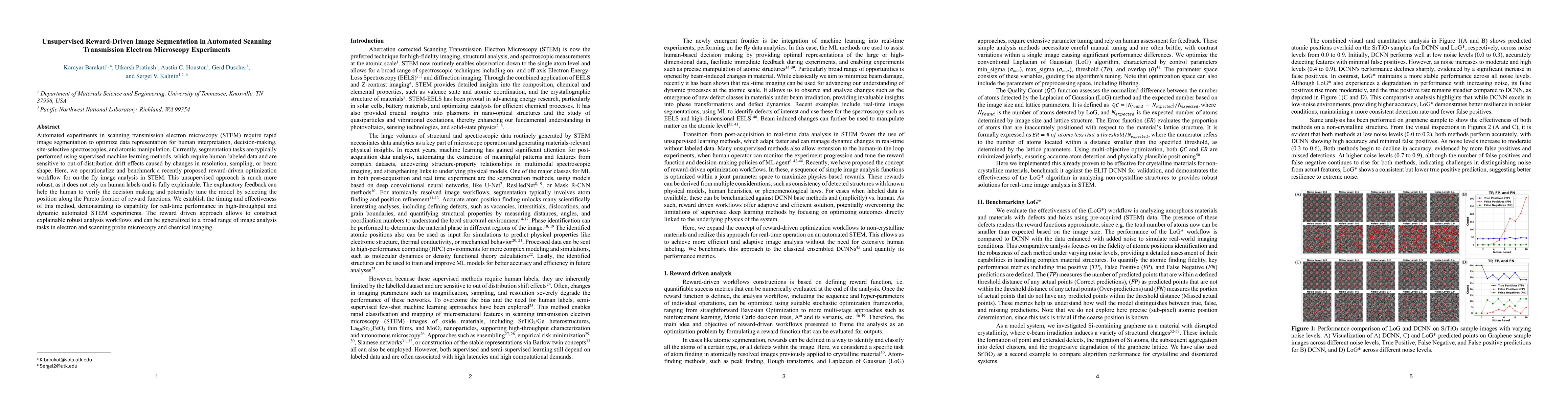
AI Key Findings

Generated Sep 02, 2025

Methodology

The research introduces a reward-driven, unsupervised workflow for real-time image segmentation in Scanning Transmission Electron Microscopy (STEM), utilizing the Laplacian of Gaussian (LoG*) method and physics-based reward functions for hyperparameter optimization. The method is benchmarked against deep learning approaches, demonstrating consistent performance across varying noise levels and structural complexities.

Key Results

  • LoG* workflow maintains consistent performance across varying noise levels and structural complexities, unlike deep learning approaches that are sensitive to out-of-distribution drift effects.
  • A sliding window approach enhances computational efficiency, enabling local optimization of parameters and reducing processing time while maintaining accuracy.
  • LoG* offers faster optimization times compared to deep learning models, making it suitable for dynamic, real-time applications.
  • The modular design of LoG* allows for easy integration of pre-processing and post-processing techniques, such as noise reduction and clustering, for effective weakly supervised defect identification.
  • The reward-based workflows enable human-in-the-loop control, allowing flexible decision-making by balancing competing objectives in real-time STEM analysis.

Significance

This research offers an unsupervised, explainable, and robust alternative to supervised methods for image segmentation in STEM, which are typically reliant on human-labeled data. The method's real-time capabilities and adaptability to changing data characteristics make it valuable for high-throughput, dynamic automated STEM experiments.

Technical Contribution

The paper presents a novel, unsupervised, reward-driven image segmentation workflow for real-time analysis in STEM experiments, utilizing LoG* with physics-based reward functions for hyperparameter optimization and a sliding window approach for computational efficiency.

Novelty

The research introduces an unsupervised, explainable, and robust image segmentation method for STEM, contrasting with supervised methods that require human-labeled data and are sensitive to drift effects. The method's real-time capabilities and adaptability to changing data characteristics distinguish it from existing approaches.

Limitations

  • The range of possible image analysis operations is more limited compared to deep learning approaches.
  • The method may not fully capture the complexity of certain image patterns that deep learning models can learn from large datasets.

Future Work

  • Extending the reward-driven concept to other microscopy techniques and imaging modalities.
  • Investigating the application of this method to other materials science and nanotechnology domains.

Paper Details

PDF Preview

Download PDF

No citations found for this paper.

Comments (0)

This website uses cookies to ensure you get the best experience. Learn more