Quick Answers
What methodology did the authors use?
The paper presents a method to convert 3D angiography images into 2D planar representations of vascular trees using a combination of classical image processing techniques, morphological filters, and a recursive embedding algorithm. The approach includes steps like graph segmentation, angular parametrization, curvature preservation, and force-directed layout refinement. More in Methodology →
What are the key results?
The method produces faithful 2D maps of vascular networks from 3D angiography data within seconds. — The planar representation preserves vessel lengths, curvature, and junction angles, which are critical for catheter navigation. More in Key Results →
Why is this work significant?
This research provides a practical tool for interventional radiology and surgery by simplifying device selection and reducing procedural risks. It also facilitates large-scale anatomical studies and improves the understanding of vascular branching patterns and tortuosity. More in Significance →
What are the main limitations?
The method may not accurately preserve small vessel geometries in 2D embeddings. — The curvature constraint is satisfied with some tolerance for smaller vessels. More in Limitations →
Abstract
The diffusion of minimally invasive, endovascular interventions motivates the development of visualization methods for complex vascular networks. We propose a planar representation of blood vessel trees which preserves the properties that are most relevant to catheter navigation: topology, length and curvature. 
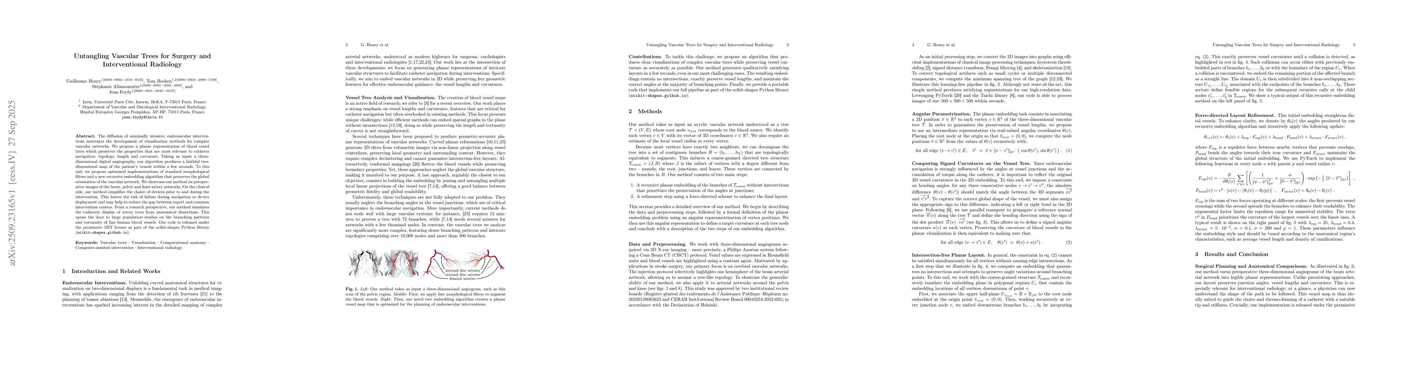
Taking as input a three-dimensional digital angiography, our algorithm produces a faithful two-dimensional map of the patient's vessels within a few seconds. To this end, we propose optimized implementations of standard morphological filters and a new recursive embedding algorithm that preserves the global orientation of the vascular network. We showcase our method on peroperative images of the brain, pelvic and knee artery networks. On the clinical side, our method simplifies the choice of devices prior to and during the intervention. This lowers the risk of failure during navigation or device deployment and may help to reduce the gap between expert and common intervention centers. From a research perspective, our method simulates the cadaveric display of artery trees from anatomical dissections. This opens the door to large population studies on the branching patterns and tortuosity of fine human blood vessels. Our code is released under the permissive MIT license as part of the scikit-shapes Python library (https://scikit-shapes.github.io ).
AI Key Findings
Generated Oct 01, 2025
Methodology — What approach did the authors take?
The paper presents a method to convert 3D angiography images into 2D planar representations of vascular trees using a combination of classical image processing techniques, morphological filters, and a recursive embedding algorithm. The approach includes steps like graph segmentation, angular parametrization, curvature preservation, and force-directed layout refinement.
Key Results — What are the main findings?
- The method produces faithful 2D maps of vascular networks from 3D angiography data within seconds.
- The planar representation preserves vessel lengths, curvature, and junction angles, which are critical for catheter navigation.
- The technique enables anatomical comparisons and supports surgical planning by visualizing detailed vascular structures.
Significance — Why does this research matter?
This research provides a practical tool for interventional radiology and surgery by simplifying device selection and reducing procedural risks. It also facilitates large-scale anatomical studies and improves the understanding of vascular branching patterns and tortuosity.
Technical Contribution — What is the technical contribution?
The paper introduces a recursive embedding algorithm that preserves the global orientation and curvature of vascular networks, combined with force-directed layout refinement for clear visualization.
Novelty — What is new about this work?
The work introduces a novel approach to planar embedding of vascular trees that maintains both geometric accuracy and readability, distinguishing it from previous topology-focused methods.
Limitations — What are the limitations of this study?
- The method may not accurately preserve small vessel geometries in 2D embeddings.
- The curvature constraint is satisfied with some tolerance for smaller vessels.
Future Work — What did the authors propose for future work?
- Extending the method to handle non-circular vessel sections.
- Integrating with state-of-the-art segmentation networks for more complex data.
- Adapting the method to vascular networks containing cycles, such as the circle of Willis.
How to Cite This Paper
@article{houry2025untangling,
title = {Untangling Vascular Trees for Surgery and Interventional Radiology},
author = {Houry, Guillaume and Feydy, Jean and Allassonnière, Stéphanie and others},
year = {2025},
eprint = {2509.23165},
archivePrefix = {arXiv},
primaryClass = {eess.IV},
doi = {10.1007/978-3-032-05114-1_64},
}
Houry, G., Feydy, J., Allassonnière, S., & Boeken, T. (2025). Untangling Vascular Trees for Surgery and Interventional Radiology. arXiv. https://doi.org/10.1007/978-3-032-05114-1_64
Houry, Guillaume, et al. "Untangling Vascular Trees for Surgery and Interventional Radiology." arXiv, 2025, doi.org/10.1007/978-3-032-05114-1_64.
PDF Preview
Similar Papers
Found 4 papersDigestive and bariatric surgery, digestive endoscopy and interventional radiology.
Kobeiter, Hicham, De Jong, Audrey, Christou, Niki et al.
Antibioprophylaxis in thoracic surgery, thoracic endoscopy, and interventional radiology.
Alifano, Marco, Ruiz, Stéphanie, Selim, Jean et al.
Emergency Preparedness in the Vascular and Interventional Radiology Suite-An Overview.
Verma, Naina, Kondoor, Vishaal, Kolyvas, Peter et al.
Imaging-Based Artificial Intelligence in Vascular and Interventional Radiology: A Narrative Review.
Wang, Ping, Ganjizadeh, Ali, Erickson, Bradley J et al.
Comments (0)