Summary
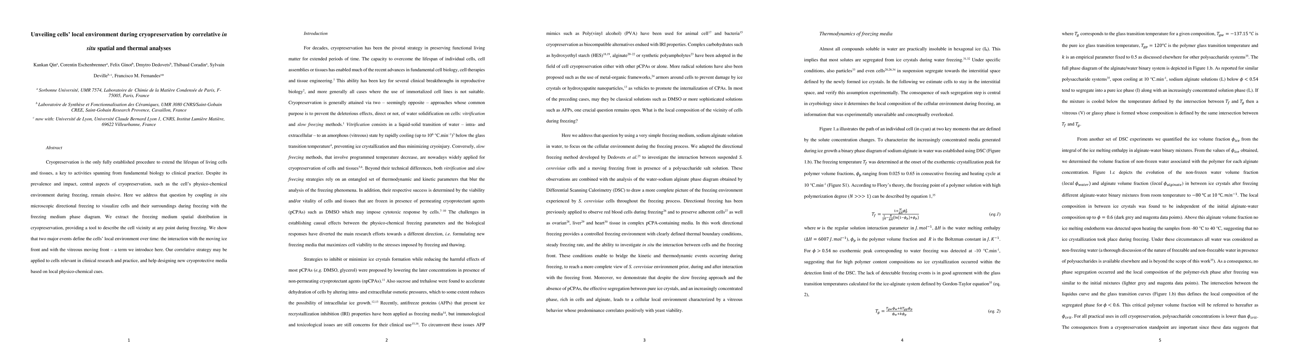
Cryopreservation is the only fully established procedure to extend the lifespan of living cells and tissues, a key to activities spanning from fundamental biology to clinical practice. Despite its prevalence and impact, central aspects of cryopreservation, such as the cell's physico-chemical environment during freezing, remain elusive. Here we address that question by coupling in situ microscopic directional freezing to visualize cells and their surroundings during freezing with the freezing medium phase diagram. We extract the freezing medium spatial distribution in cryopreservation, providing a tool to describe the cell vicinity at any point during freezing. We show that two major events define the cells' local environment over time: the interaction with the moving ice front and with the vitreous moving front - a term we introduce here. Our correlative strategy may be applied to cells relevant in clinical research and practice, and help designing new cryoprotective media based on local physico-chemical cues.
AI Key Findings
Get AI-generated insights about this paper's methodology, results, and significance.
Paper Details
PDF Preview
Key Terms
Citation Network
Current paper (gray), citations (green), references (blue)
Display is limited for performance on very large graphs.
Similar Papers
Found 4 papersCryopreservation of siRNA-treated cells is feasible
Sauer, M., Echeverria, D., Kremer, A. et al.
Correlative and in situ microscopy investigation of phase transformation, crystal growth and degradation of antimony sulfide thin films
Johannes Will, Janina Maultzsch, Tobias Dierke et al.
No citations found for this paper.
Comments (0)