01
MethodologyHow they did it
Structured illumination microscopy using upconversion nanoparticles for super-resolution imaging
Researchers developed a new microscopy technique that uses upconversion to visualize biomolecules inside cells at high resolution, surpassing the limitations of traditional structured illumination microscopy through biological tissues.
Researchers developed a new microscopy technique that uses upconversion to visualize biomolecules inside cells at high resolution, surpassing the limitations of traditional structured illumination microscopy through biological tissues.
Structured illumination microscopy using upconversion nanoparticles for super-resolution imaging More in Methodology →
Main finding 1: Upconversion nanoparticles enable high-resolution imaging of cellular structures at nanoscale resolution. — Main finding 2: The method is suitable for imaging thick samples and can track single nanoparticles in living cells. More in Key Results →
This research contributes to the development of super-resolution microscopy and its applications in biological and material sciences. More in Significance →
Limitation 1: The method requires high-quality upconversion nanoparticles and can be affected by sample quality. — Limitation 2: The technique is sensitive to photobleaching and requires optimization for long-term imaging. More in Limitations →
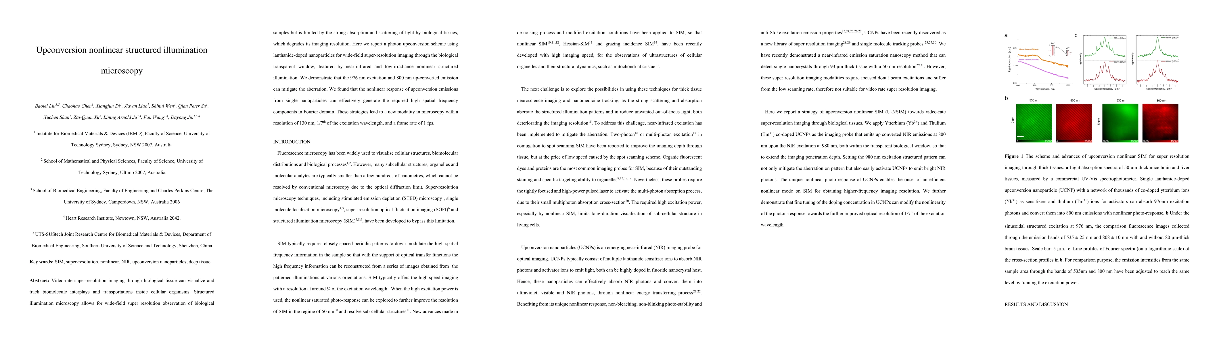
Video-rate super-resolution imaging through biological tissue can visualize and track biomolecule interplays and transportations inside cellular organisms. Structured illumination microscopy allows for wide-field super resolution observation of biological samples but is limited by the strong absorption and scattering of light by biological tissues, which degrades its imaging resolution. Here we report a photon upconversion scheme using lanthanide-doped nanoparticles for wide-field super-resolution imaging through the biological transparent window, featured by near-infrared and low-irradiance nonlinear structured illumination. We demonstrate that the 976 nm excitation and 800 nm up-converted emission can mitigate the aberration. We found that the nonlinear response of upconversion emissions from single nanoparticles can effectively generate the required high spatial frequency components in Fourier domain. These strategies lead to a new modality in microscopy with a resolution of 130 nm, 1/7th of the excitation wavelength, and a frame rate of 1 fps.
Seven facets of this paper, analysed and brought into focus by AI.
This research contributes to the development of super-resolution microscopy and its applications in biological and material sciences.
Structured illumination microscopy using upconversion nanoparticles for super-resolution imaging
This research contributes to the development of super-resolution microscopy and its applications in biological and material sciences.
The development of a novel method for super-resolution imaging using upconversion nanoparticles.
This work presents a new approach to super-resolution microscopy that combines the benefits of structured illumination with the unique properties of upconversion nanoparticles.
Current paper (gray), citations (green), references (blue)
Display is limited for performance on very large graphs.
Discussion 0