Upconverting microgauges reveal intraluminal force dynamics in vivo

PDF

Publication

Published: Oct 09, 2025
Categories: q-bio.TO, q-bio.QM

Metrics

Source: ArXiv

Quick Actions

View PDF

Quick Answers

What methodology did the authors use?

The study employed a combination of upconversion luminescence (UCL) spectroscopy, diamond anvil cell (DAC) experiments, and electrophysiological recordings to investigate pressure-induced changes in microgauge luminescence and their correlation with pharyngeal function. More in Methodology →

What are the key results?

High pressure induces a shift in Er3+ upconversion luminescence color from green to red, attributed to cross-relaxation and matrix quenching mechanisms. — Polystyrene matrix quenching enhances the IRed:IGreen ratio at elevated pressures due to pressure-induced blueshifting of vibrational modes and improved resonance with green quenching transitions. More in Key Results →

Why is this work significant?

This research provides critical insights into pressure-induced optical phenomena in upconversion nanophosphors and their potential applications in biophotonics, pressure sensing, and mechanosensitive biological systems. More in Significance →

What are the main limitations?

The study relies on indirect measurements of pressure effects through luminescence shifts, which may not fully capture all underlying physical mechanisms. — The use of polystyrene as a matrix may limit generalizability to other biological or synthetic environments. More in Limitations →

Paper Preview

Abstract

The forces generated by action potentials in muscle cells shuttle blood, food and waste products throughout the luminal structures of the body. Although non-invasive electrophysiological techniques exist, most mechanosensors cannot access luminal structures non-invasively. Here we introduce non-toxic ingestible mechanosensors to enable the quantitative study of luminal forces and apply them to study feeding in living Caenorhabditis elegans roundworms. 
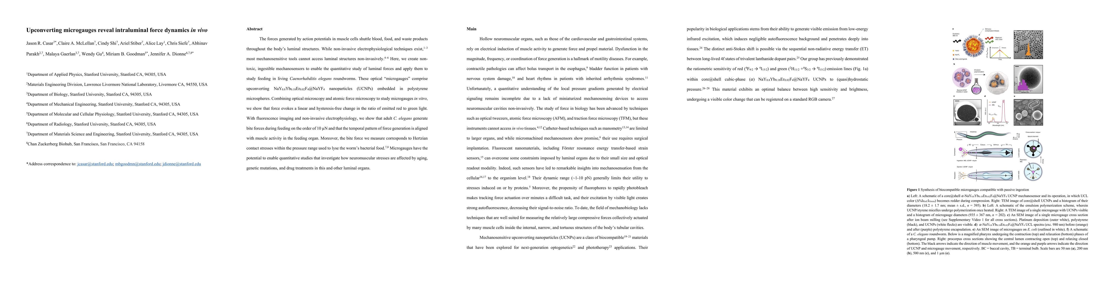
These optical 'microgauges' comprise NaY0.8Yb0.18Er0.02F4@NaYF4 upconverting nanoparticles embedded in polystyrene microspheres. Combining optical microscopy and atomic force microscopy to study microgauges in vitro, we show that force evokes a linear and hysteresis-free change in the ratio of emitted red to green light. With fluorescence imaging and non-invasive electrophysiology, we show that adult C. elegans generate bite forces during feeding on the order of 10 micronewtons and that the temporal pattern of force generation is aligned with muscle activity in the feeding organ. Moreover, the bite force we measure corresponds to Hertzian contact stresses in the pressure range used to lyse the bacterial food of the worm. Microgauges have the potential to enable quantitative studies that investigate how neuromuscular stresses are affected by aging, genetic mutations and drug treatments in this organ and other luminal organs.

AI Key Findings

Generated Oct 11, 2025

Methodology — What approach did the authors take?

The study employed a combination of upconversion luminescence (UCL) spectroscopy, diamond anvil cell (DAC) experiments, and electrophysiological recordings to investigate pressure-induced changes in microgauge luminescence and their correlation with pharyngeal function.

Key Results — What are the main findings?

  • High pressure induces a shift in Er3+ upconversion luminescence color from green to red, attributed to cross-relaxation and matrix quenching mechanisms.
  • Polystyrene matrix quenching enhances the IRed:IGreen ratio at elevated pressures due to pressure-induced blueshifting of vibrational modes and improved resonance with green quenching transitions.
  • Pharyngeal muscle activity was successfully synchronized with microgauge luminescence data, revealing pressure-dependent correlations between luminescence changes and physiological responses.

Significance — Why does this research matter?

This research provides critical insights into pressure-induced optical phenomena in upconversion nanophosphors and their potential applications in biophotonics, pressure sensing, and mechanosensitive biological systems.

Technical Contribution — What is the technical contribution?

The work establishes a novel framework for correlating pressure-induced optical changes in upconversion nanophosphors with physiological responses, integrating UCL spectroscopy, DAC experiments, and electrophysiology.

Novelty — What is new about this work?

This study uniquely combines UCL spectroscopy with electrophysiological recordings to demonstrate pressure-dependent luminescence shifts and their physiological implications, offering new perspectives on mechanosensitive optical sensing in biological systems.

Limitations — What are the limitations of this study?

  • The study relies on indirect measurements of pressure effects through luminescence shifts, which may not fully capture all underlying physical mechanisms.
  • The use of polystyrene as a matrix may limit generalizability to other biological or synthetic environments.

Future Work — What did the authors propose for future work?

  • Investigate pressure-dependent quenching mechanisms in alternative matrices such as silica or biological tissues.
  • Develop high-resolution pressure-sensing devices based on microgauge luminescence shifts.
  • Explore the role of local symmetry and refractive index changes in pressure-induced UCL modulation.

Paper Details

Paper ID: 2510.07657
Comments: 18 pages, 4 figures
Categories:
q-bio.TO q-bio.QM

How to Cite This Paper

BibTeX
@article{parakh2025upconverting,
  title         = {Upconverting microgauges reveal intraluminal force dynamics in vivo},
  author        = {Parakh, Abhinav and Lay, Alice and Stiber, Ariel and others},
  year          = {2025},
  eprint        = {2510.07657},
  archivePrefix = {arXiv},
  primaryClass  = {q-bio.TO},
  doi           = {10.1038/s41586-024-08331-x},
}
APA 7
Parakh, A., Lay, A., Stiber, A., Siefe, C., Shi, C., McLellan, C., Casar, J., Dionne, J., Gaerlan, M., Goodman, M., & Gu, X. (2025). Upconverting microgauges reveal intraluminal force dynamics in vivo. arXiv. https://doi.org/10.1038/s41586-024-08331-x
MLA 9
Parakh, Abhinav, et al. "Upconverting microgauges reveal intraluminal force dynamics in vivo." arXiv, 2025, doi.org/10.1038/s41586-024-08331-x.

Comments (0)

This website uses cookies to ensure you get the best experience. Learn more