ArxivLens

Quick Summary:

This paper proposes an algorithm for left ventricular segmentation in long axis ultrasound images by first detecting anatomical markers, such as the mitral valve and apex, to create an initial contour guess. The method shows good agreement with manually traced contours, indicating reliable left ventricle border detection.

arXiv Id: 1510.03250
Comments: 11 pages, 17 figures
Date Published: 2015-10-13
Date Updated: 2015-10-13

Categories:

Key Terms (TF-IDF):

left (0.299)
left (0.295)
segmentation (0.271)
segmentation (0.251)
initial (0.241)
detection (0.239)
initial (0.236)
algorithm (0.236)
detection (0.231)
algorithm (0.229)
ultrasound (0.204)
passing (0.184)
passing (0.178)
manually (0.177)
creating (0.173)
indices (0.173)
indices (0.169)
automatic (0.161)
creating (0.160)
detecting (0.158)
License: http://arxiv.org/licenses/nonexclusive-distrib/1.0/

Summary:

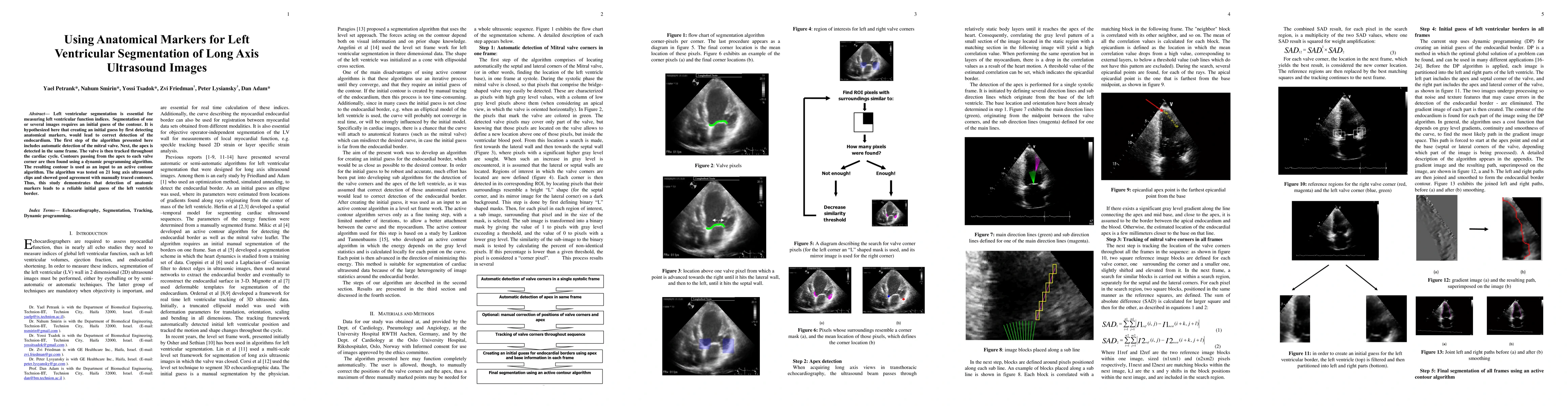
Left ventricular segmentation is essential for measuring left ventricular function indices. Segmentation of one or several images requires an initial guess of the contour. It is hypothesized here that creating an initial guess by first detecting anatomical markers, would lead to correct detection of the endocardium. The first step of the algorithm presented here includes automatic detection of the mitral valve. Next, the apex is detected in the same frame. The valve is then tracked throughout the cardiac cycle. Contours passing from the apex to each valve corner are then found using a dynamic programming algorithm. The resulting contour is used as an input to an active contour algorithm. The algorithm was tested on 21 long axis ultrasound clips and showed good agreement with manually traced contours. Thus, this study demonstrates that detection of anatomic markers leads to a reliable initial guess of the left ventricle border.

Citation & Reference Network Visualization

This graph shows how this paper is connected to other research. The current paper is shown in dark gray, papers that cite it are in green, and papers it references are in blue. You can:

Current Paper
Papers citing this work (Citations)
Papers cited by this work (References)

• Hover over any node to see the paper title

• Click a node to view that paper's details

• Drag nodes to rearrange the graph

• Use the zoom controls (bottom right) to zoom in/out

Citations and References

Title Authors Year Actions
Blood flow patterns estimation in the left ventricle with low-rate 2D and 3D dynamic contrast-enhanced ultrasound
Peiran Chen, R. V. Sloun, S. Turco, H. Wijkstra, D. Filomena, L. Agati, P. Houthuizen, M. Mischi
2020
View Paper
Dynamic velocity vector and relative pressure estimation in the left ventricle with dynamic contrast-enhanced ultrasound of low frame rates
Peiran Chen, R. V. Sloun, S. Turco, H. Wijkstra, P. Houthuizen, M. Mischi
2018
View Paper
Optimization-Based Speckle Tracking Algorithm for Left Ventricle Strain Estimation: A Feasibility Study
H. Khamis, S. Shimoni, A. Hagendorff, N. Smirin, Z. Friedman, D. Adam
2016
View Paper

Comments (0)

This website uses cookies to ensure you get the best experience. Learn more