Publication
Metrics
AI Quick Summary
This study develops a computer-aided diagnosis system using a Spatio-Temporal Dual-Stream Network with self-supervised learning to classify benign and malignant lung lesions from radial probe endobronchial ultrasound videos. The proposed method enhances noise robustness and achieves high accuracy, reducing the need for manual image selection and improving real-time diagnostic efficiency.
Paper Preview
Abstract
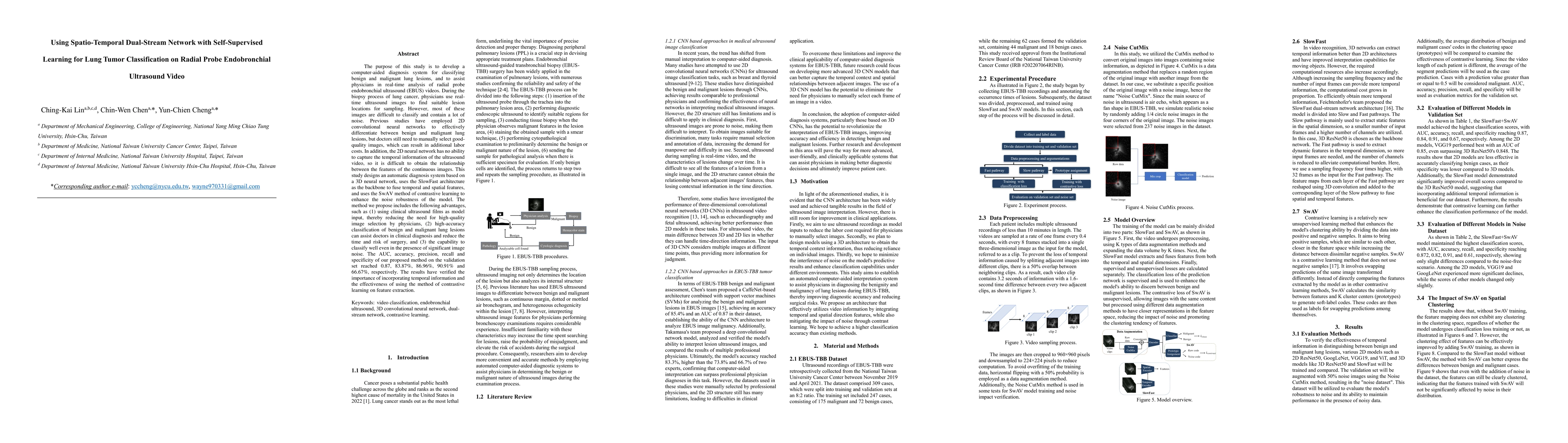
The purpose of this study is to develop a computer-aided diagnosis system for classifying benign and malignant lung lesions, and to assist physicians in real-time analysis of radial probe endobronchial ultrasound (EBUS) videos. During the biopsy process of lung cancer, physicians use real-time ultrasound images to find suitable lesion locations for sampling. However, most of these images are difficult to classify and contain a lot of noise. Previous studies have employed 2D convolutional neural networks to effectively differentiate between benign and malignant lung lesions, but doctors still need to manually select good-quality images, which can result in additional labor costs. In addition, the 2D neural network has no ability to capture the temporal information of the ultrasound video, so it is difficult to obtain the relationship between the features of the continuous images. This study designs an automatic diagnosis system based on a 3D neural network, uses the SlowFast architecture as the backbone to fuse temporal and spatial features, and uses the SwAV method of contrastive learning to enhance the noise robustness of the model. The method we propose includes the following advantages, such as (1) using clinical ultrasound films as model input, thereby reducing the need for high-quality image selection by physicians, (2) high-accuracy classification of benign and malignant lung lesions can assist doctors in clinical diagnosis and reduce the time and risk of surgery, and (3) the capability to classify well even in the presence of significant image noise. The AUC, accuracy, precision, recall and specificity of our proposed method on the validation set reached 0.87, 83.87%, 86.96%, 90.91% and 66.67%, respectively. The results have verified the importance of incorporating temporal information and the effectiveness of using the method of contrastive learning on feature extraction.
AI Key Findings
Get AI-generated insights about this paper's methodology, results, significance, and more — seven facets brought into focus.
Impact
Paper Details
Authors
PDF Preview
Key Terms
Citation Network
Current paper (gray), citations (green), references (blue)
Display is limited for performance on very large graphs.
Discussion 0