Using the Polar Transform for Efficient Deep Learning-Based Aorta Segmentation in CTA Images
Publication
Metrics
AI Quick Summary
Their approach uses polar image transformations and hysteresis thresholding to fuse predictions, outperforming state-of-the-art methods while maintaining pixel-level recall.
Paper Preview
Abstract
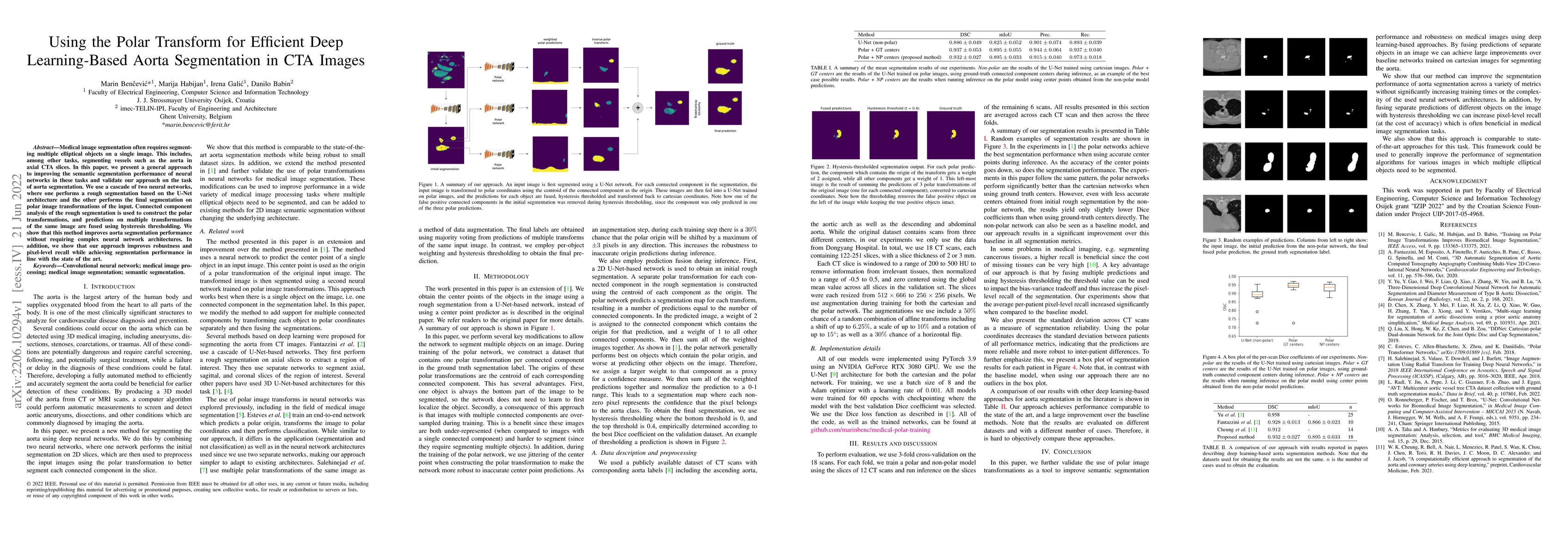
Medical image segmentation often requires segmenting multiple elliptical objects on a single image. This includes, among other tasks, segmenting vessels such as the aorta in axial CTA slices. In this paper, we present a general approach to improving the semantic segmentation performance of neural networks in these tasks and validate our approach on the task of aorta segmentation. We use a cascade of two neural networks, where one performs a rough segmentation based on the U-Net architecture and the other performs the final segmentation on polar image transformations of the input. Connected component analysis of the rough segmentation is used to construct the polar transformations, and predictions on multiple transformations of the same image are fused using hysteresis thresholding. We show that this method improves aorta segmentation performance without requiring complex neural network architectures. In addition, we show that our approach improves robustness and pixel-level recall while achieving segmentation performance in line with the state of the art.
AI Key Findings
Get AI-generated insights about this paper's methodology, results, significance, and more — seven facets brought into focus.
Impact
Paper Details
Authors
PDF Preview
Key Terms
Citation Network
Current paper (gray), citations (green), references (blue)
Display is limited for performance on very large graphs.
Discussion 0