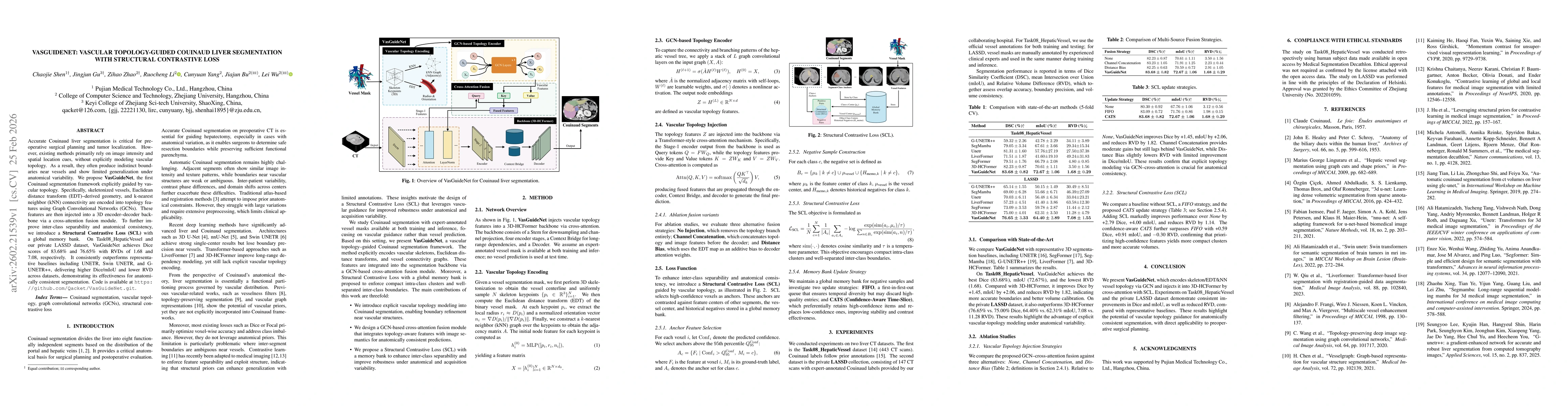
VasGuideNet encodes vessel skeletons, Euclidean distance transforms, and k‑nearest‑neighbor graphs with a GCN, injects the resulting topology features into a 3D‑HCFormer backbone via cross‑attention, and trains with a structural contrastive loss that uses a global memory bank.
VasGuideNet: Vascular Topology-Guided Couinaud Liver Segmentation with Structural Contrastive Loss
Publication
Metrics
Quick Answers
What methodology did the authors use?
VasGuideNet encodes vessel skeletons, Euclidean distance transforms, and k‑nearest‑neighbor graphs with a GCN, injects the resulting topology features into a 3D‑HCFormer backbone via cross‑attention, and trains with a structural contrastive loss that uses a global memory bank. More in Methodology →
What are the key results?
Dice 83.68% on Task08_HepaticVessel and 76.65% on LASSD, surpassing all baselines — mIoU improved by 2–3% and RVD reduced to 1.68% on Task08, 7.08% on LASSD More in Key Results →
Why is this work significant?
The method delivers anatomically consistent liver segmentation, especially near vessels, and improves robustness to anatomical variability, which is critical for surgical planning. More in Significance →
What are the main limitations?
Requires expert‑annotated vessel masks at training and inference — Computational overhead from 3D skeletonization and GCN may limit real‑time deployment More in Limitations →
Abstract
Accurate Couinaud liver segmentation is critical for preoperative surgical planning and tumor localization.However, existing methods primarily rely on image intensity and spatial location cues, without explicitly modeling vascular topology. As a result, they often produce indistinct boundaries near vessels and show limited generalization under anatomical variability.We propose VasGuideNet, the first Couinaud segmentation framework explicitly guided by vascular topology. Specifically, skeletonized vessels, Euclidean distance transform (EDT)--derived geometry, and k-nearest neighbor (kNN) connectivity are encoded into topology features using Graph Convolutional Networks (GCNs). These features are then injected into a 3D encoder--decoder backbone via a cross-attention fusion module. To further improve inter-class separability and anatomical consistency, we introduce a Structural Contrastive Loss (SCL) with a global memory bank.On Task08_HepaticVessel and our private LASSD dataset, VasGuideNet achieves Dice scores of 83.68% and 76.65% with RVDs of 1.68 and 7.08, respectively. It consistently outperforms representative baselines including UNETR, Swin UNETR, and G-UNETR++, delivering higher Dice/mIoU and lower RVD across datasets, demonstrating its effectiveness for anatomically consistent segmentation. Code is available at https://github.com/Qacket/VasGuideNet.git.
Key Findings, in focus
Seven facets of this paper, analysed and brought into focus by AI.
The method delivers anatomically consistent liver segmentation, especially near vessels, and improves robustness to anatomical variability, which is critical for surgical planning.
- Dice 83.68% on Task08_HepaticVessel and 76.65% on LASSD, surpassing all baselines
- mIoU improved by 2–3% and RVD reduced to 1.68% on Task08, 7.08% on LASSD
- Ablation shows GCN‑cross‑attention and CATS memory update yield the best performance
The method delivers anatomically consistent liver segmentation, especially near vessels, and improves robustness to anatomical variability, which is critical for surgical planning.
A novel GCN‑based cross‑attention fusion of vascular topology into a transformer backbone, coupled with a structural contrastive loss that enforces inter‑class separability using a memory bank.
First explicit use of vascular topology for Couinaud liver segmentation, combining graph convolution, cross‑attention, and contrastive learning in a unified framework.
- Requires expert‑annotated vessel masks at training and inference
- Computational overhead from 3D skeletonization and GCN may limit real‑time deployment
- Develop vessel‑prediction modules to remove the need for manual vessel masks
- Apply the topology‑guided framework to other organs or imaging modalities
Discussion 0