Ventricular Segmentation: A Brief Comparison of U-Net Derivatives
Publication
Metrics
AI Quick Summary
This paper compares various U-Net derivative architectures for the semantic segmentation of cardiac MRI images, aiming to improve heart disorder diagnosis and treatment. It evaluates the models' efficacy through quantitative metrics and discusses challenges and future improvement strategies.
Paper Preview
Abstract
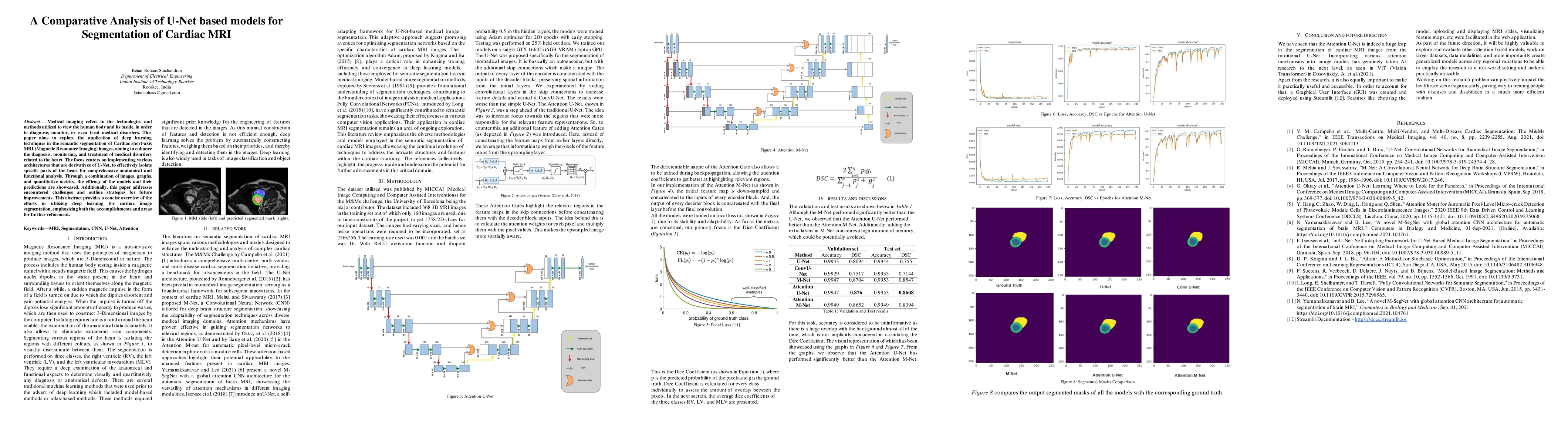
Medical imaging refers to the technologies and methods utilized to view the human body and its inside, in order to diagnose, monitor, or even treat medical disorders. This paper aims to explore the application of deep learning techniques in the semantic segmentation of Cardiac short-axis MRI (Magnetic Resonance Imaging) images, aiming to enhance the diagnosis, monitoring, and treatment of medical disorders related to the heart. The focus centers on implementing various architectures that are derivatives of U-Net, to effectively isolate specific parts of the heart for comprehensive anatomical and functional analysis. Through a combination of images, graphs, and quantitative metrics, the efficacy of the models and their predictions are showcased. Additionally, this paper addresses encountered challenges and outline strategies for future improvements. This abstract provides a concise overview of the efforts in utilizing deep learning for cardiac image segmentation, emphasizing both the accomplishments and areas for further refinement.
AI Key Findings
Get AI-generated insights about this paper's methodology, results, significance, and more — seven facets brought into focus.
Impact
Paper Details
Authors
PDF Preview
Key Terms
Citation Network
Current paper (gray), citations (green), references (blue)
Display is limited for performance on very large graphs.
Discussion 0