Summary
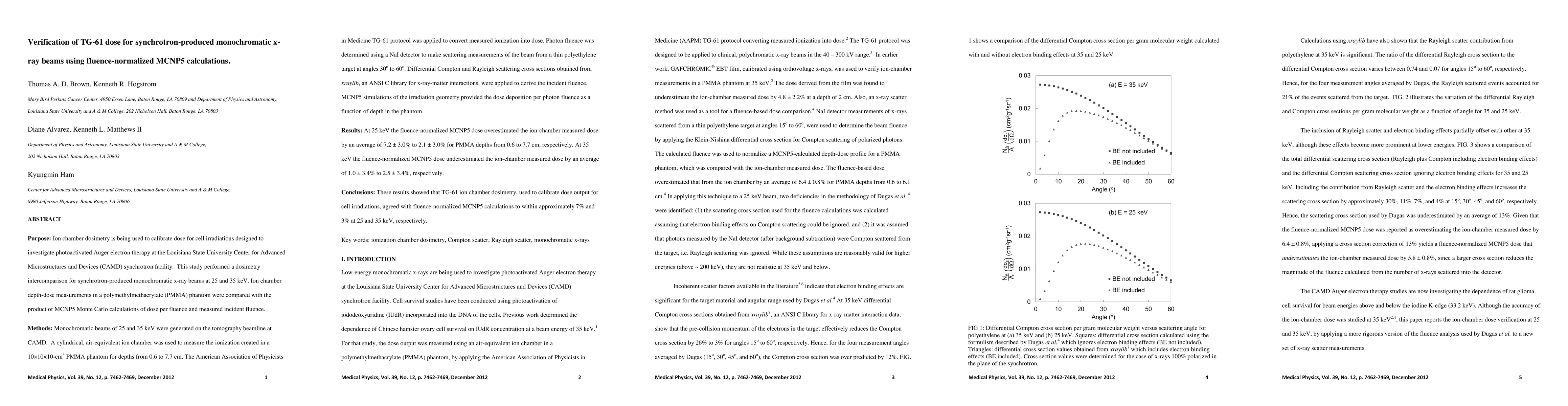
Ion chamber dosimetry is being used to calibrate dose for cell irradiations designed to investigate photoactivated Auger electron therapy at the Louisiana State University CAMD synchrotron facility. This study performed a dosimetry intercomparison for synchrotron-produced monochromatic x-ray beams at 25 and 35 keV. Ion chamber depth-dose measurements in a PMMA phantom were compared with the product of MCNP5 Monte Carlo calculations of dose per fluence and measured incident fluence. Monochromatic beams of 25 and 35 keV were generated on the tomography beamline at CAMD. A cylindrical, air-equivalent ion chamber was used to measure the ionization created in a 10x10x10-cm3 PMMA phantom for depths from 0.6 to 7.7 cm. The American Association of Physicists in Medicine TG-61 protocol was applied to convert measured ionization into dose. Photon fluence was determined using a NaI detector to make scattering measurements of the beam from a thin polyethylene target at angles 30 degrees to 60 degrees. Differential Compton and Rayleigh scattering cross sections obtained from xraylib, an ANSI C library for x-ray-matter interactions, were applied to derive the incident fluence. MCNP5 simulations of the irradiation geometry provided the dose deposition per photon fluence as a function of depth in the phantom. At 25 keV the fluence-normalized MCNP5 dose overestimated the ion-chamber measured dose by an average of 7.2+/-3.0% to 2.1+/-3.0% for PMMA depths from 0.6 to 7.7 cm, respectively. At 35 keV the fluence-normalized MCNP5 dose underestimated the ion-chamber measured dose by an average of 1.0+/-3.4% to 2.5+/-3.4%, respectively. These results showed that TG-61 ion chamber dosimetry, used to calibrate dose output for cell irradiations, agreed with fluence-normalized MCNP5 calculations to within approximately 7% and 3% at 25 and 35 keV, respectively.
AI Key Findings
Generated Sep 07, 2025
Methodology
The study performed a dosimetry intercomparison for synchrotron-produced monochromatic x-ray beams at 25 and 35 keV using fluence-normalized MCNP5 calculations, comparing ion chamber depth-dose measurements in a PMMA phantom with MCNP5-calculated dose per fluence and measured incident fluence.
Key Results
- Fluence-normalized MCNP5 dose overestimated ion-chamber measured dose by an average of 7.2±3.0% to 2.1±3.0% for PMMA depths from 0.6 to 7.7 cm at 25 keV.
- Fluence-normalized MCNP5 dose underestimated ion-chamber measured dose by an average of 1.0±3.4% to 2.5±3.4% for PMMA depths from 0.6 to 7.7 cm at 35 keV.
- TG-61 ion chamber dosimetry agreed with fluence-normalized MCNP5 calculations to within approximately 7% and 3% at 25 and 35 keV, respectively.
Significance
This research is important for verifying the AAPM TG-61 ion chamber dosimetry used to calibrate dose output from monochromatic x-ray beams for photoactivated Auger electron therapy, ensuring accurate dose calibration for cell irradiation experiments.
Technical Contribution
The study improved upon previous work by revising Compton cross section calculations to include electron binding effects and incorporating the Rayleigh scatter contribution into fluence calculations.
Novelty
This work presents significant improvements over previous dosimetry intercomparison methods, specifically in the accuracy of fluence-MCNP5 dose distribution for monochromatic x-ray beams.
Limitations
- The study was limited by uncertainties in broad beam fluence, including counting statistics, beam output fluctuations, solid angle subtended by the detector collimator aperture, and uncertainty in the width of the collimated narrow beam.
- The low number of n=2 photons contributed small uncertainty to the total dose uncertainty.
Future Work
- Further research could benefit from using an additional dose measurement device, such as a calorimeter, and extending TG-61 to include monochromatic x-ray beams.
- Investigating the stability of the high-energy region of the photon spectrum beyond 35 keV may improve dose measurement accuracy.
Paper Details
PDF Preview
Key Terms
Citation Network
Current paper (gray), citations (green), references (blue)
Display is limited for performance on very large graphs.
Similar Papers
Found 4 papersFluence Adaptation for Task-based Dose Optimization in X-ray Phase-Contrast Imaging
Xi Zhang, Li Zhang, Yuxiang Xing et al.
| Title | Authors | Year | Actions |
|---|
Comments (0)