Virtual Alignment Method and its application to the dental prostheses and diagnosis
Publication
Metrics
AI Quick Summary
The Virtual Alignment Method (VAM) enhances the accuracy of X-ray tomography by minimizing errors, particularly in dental imaging where patient movement is a significant issue. The method's application to dental prostheses and diagnostics shows promise in producing high-quality images and precise reconstructions.
Paper Preview
Abstract
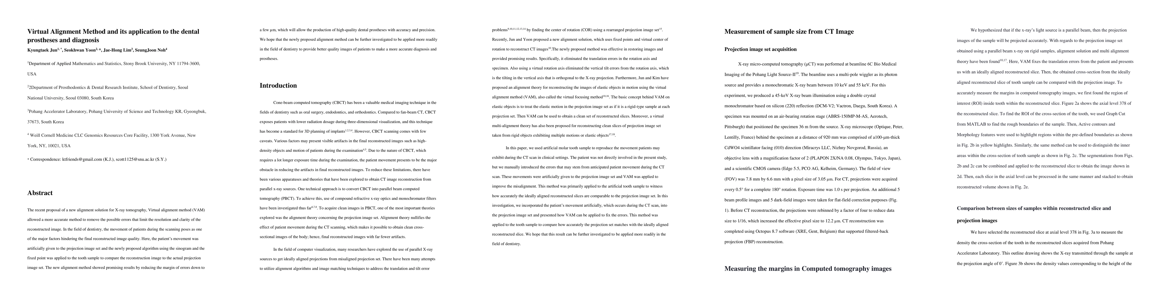
The recent proposal of a new alignment solution for X-ray tomography, Virtual alignment method (VAM) allowed a more accurate method to remove the possible errors that limit the resolution and clarity of the reconstructed image. In the field of dentistry, the movement of patients during the scanning poses as one of the major factors hindering the final reconstructed image quality. Here, the patient's movement was artificially given to the projection image set and the newly proposed algorithm using the sinogram and the fixed point was applied to the tooth sample to compare the reconstruction image to the actual projection image set. The new alignment method showed promising results by reducing the margin of errors down to a few micrometer, which will allow the production of high-quality dental prostheses with accuracy and precision. We hope that the newly proposed alignment method can be further investigated to be applied more readily in the filed of dentistry ot provide better quality images of patients to make a more accurate diagnosis and prostheses.
AI Key Findings
Get AI-generated insights about this paper's methodology, results, significance, and more — seven facets brought into focus.
Impact
Paper Details
Authors
PDF Preview
Key Terms
Citation Network
Current paper (gray), citations (green), references (blue)
Display is limited for performance on very large graphs.
Discussion 0