ArxivLens

Quick Summary:

This paper introduces a deep-learning-based method for virtual histochemical staining of label-free total-absorption photoacoustic remote sensing (TA-PARS) images, using a Pix2Pix GAN to generate H&E-like visualizations from unstained tissue. Dermatologic pathologists confirmed the diagnostic quality of the virtual stains, suggesting a potential reduction in time-to-histological imaging.

arXiv Id: 2203.02584
Comments: 16 pages, 8 figures
Date Published: 2022-04-04
Date Updated: 2022-04-04

Key Terms (TF-IDF):

tissue (0.258)
label (0.221)
label (0.220)
images (0.210)
images (0.208)
assessment (0.196)
virtual (0.188)
assessment (0.185)
radiative (0.182)
virtual (0.179)
free (0.172)
free (0.172)
radiative (0.172)
absorption (0.160)
absorption (0.157)
need (0.143)
need (0.140)
total (0.137)
skin (0.135)
remote sensing (0.133)
License: http://creativecommons.org/licenses/by/4.0/

Summary:

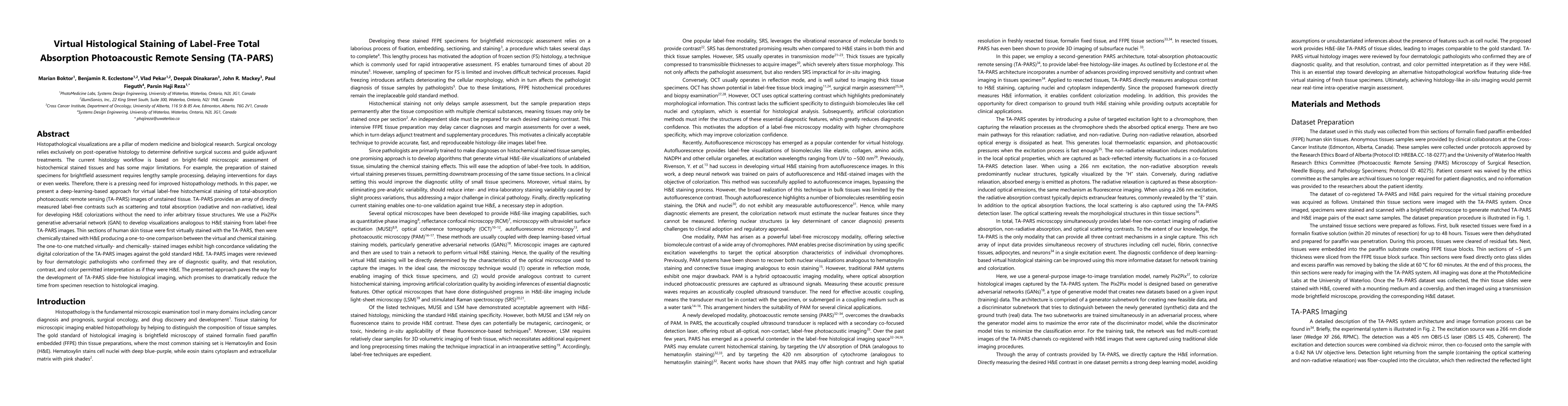
Histopathological visualizations are a pillar of modern medicine and biological research. Surgical oncology relies exclusively on post-operative histology to determine definitive surgical success and guide adjuvant treatments. The current histology workflow is based on bright-field microscopic assessment of histochemical stained tissues and has some major limitations. For example, the preparation of stained specimens for brightfield assessment requires lengthy sample processing, delaying interventions for days or even weeks. Hence, there is a pressing need for improved histopathology methods. In this paper, we present a deep-learning-based approach for virtual label-free histochemical staining of total-absorption photoacoustic remote sensing (TA-PARS) images of unstained tissue. TA-PARS provides an array of directly measured label-free contrasts such as scattering and total absorption (radiative and non-radiative), ideal for developing H&E colorizations without the need to infer arbitrary tissue structures. We use a Pix2Pix generative adversarial network (GAN) to develop visualizations analogous to H&E staining from label-free TA-PARS images. Thin sections of human skin tissue were first virtually stained with the TA-PARS, then were chemically stained with H&E producing a one-to-one comparison between the virtual and chemical staining. The one-to-one matched virtually- and chemically- stained images exhibit high concordance validating the digital colorization of the TA-PARS images against the gold standard H&E. TA-PARS images were reviewed by four dermatologic pathologists who confirmed they are of diagnostic quality, and that resolution, contrast, and color permitted interpretation as if they were H&E. The presented approach paves the way for the development of TA-PARS slide-free histology, which promises to dramatically reduce the time from specimen resection to histological imaging.

Citation & Reference Network Visualization

This graph shows how this paper is connected to other research. The current paper is shown in dark gray, papers that cite it are in green, and papers it references are in blue. You can:

Current Paper
Papers citing this work (Citations)
Papers cited by this work (References)

• Hover over any node to see the paper title

• Click a node to view that paper's details

• Drag nodes to rearrange the graph

• Use the zoom controls (bottom right) to zoom in/out

Citations and References

Title Authors Year Actions
Simultaneous Ultraviolet Photoacoustic and Auto‐Fluorescence Microscopy for High‐Contrast Histological Imaging
Xiufeng Li, Bingxin Huang, Claudia T. K. Lo, Chengbo Liu, Terence T. W. Wong
2025
View Paper
Clinical translation of photoacoustic imaging
Jeongwoo Park, Seongwook Choi, F. Knieling, Bryan Clingman, Sarah E Bohndiek, Lihong V. Wang, Chulhong Kim
2024
View Paper
Deep learning-based virtual staining, segmentation, and classification in label-free photoacoustic histology of human specimens
Chiho Yoon, Eunwoo Park, Sampa Misra, J. Kim, J. Baik, Kwang Gi Kim, Chan Kwon Jung, Chulhong Kim
2024
View Paper
Evaluation of tumor budding with virtual panCK stains generated by novel multi-model CNN framework.
Xingzhong Hou, Zhen Guan, Xianwei Zhang, Xiao Hu, S. Zou, Chunzi Liang, Lulin Shi, Kaitai Zhang, Haihang You
2024
View Paper
Virtual Gram staining of label-free bacteria using dark-field microscopy and deep learning
Çaǧatay Işıl, H. Koydemir, Merve Eryilmaz, Kevin de Haan, N. Pillar, Koray Mentesoglu, Aras Firat Unal, Y. Rivenson, Sukantha Chandrasekaran, O. Garner, Aydogan Ozcan
2024
View Paper
Virtual Immunohistochemistry Staining for Histological Images Assisted by Weakly-supervised Learning
Jiahan Li, Jiuyang Dong, Shenjin Huang, Xi Li, Junjun Jiang, Xiaopeng Fan, Yongbing Zhang
2024
View Paper
Single color digital H&E staining with In-and-Out Net
Mengkun Chen, Yen-Tung Liu, Fadeel Sher Khan, Matthew C. Fox, J. Reichenberg, F. C. Lopes, Katherine R. Sebastian, Mia K. Markey, James W. Tunnell
2024
View Paper
Fast and label-free 3D virtual H&E histology via active modulation-assisted dynamic full-field OCT
Zichen Yin, Bin He, Yuzhe Ying, Shuwei Zhang, Panqi Yang, Zhengyu Chen, Zhangwei Hu, Yejiong Shi, Ruizhi Xue, Chengming Wang, Shu Wang, Guihuai Wang, Ping Xue
2024
View Paper
Dual contrastive learning based image-to-image translation of unstained skin tissue into virtually stained H&E images
Muhammad Zeeshan Asaf, Babar K Rao, Muhammad Usman Akram, S. G. Khawaja, Samavia Khan, T. Truong, Palveen Sekhon, Irfan J Khan, Muhammad Shahmir Abbasi
2024
View Paper
Metabolic light absorption, scattering, and emission (MetaLASE) microscopy
Brendon S. Restall, Nathaniel J. M. Haven, Matthew T Martell, Brendyn D. Cikaluk, Joy Wang, Pradyumna Kedarisetti, Saymon Tejay, Benjamin A. Adam, G. Sutendra, Xingyu Li, Roger J. Zemp
2024
View Paper
An end-to-end workflow for nondestructive 3D pathology.
K. Bishop, Lindsey A. Erion Barner, Qinghua Han, Elena Baraznenok, Lydia Lan, Chetan Poudel, Gan Gao, R. Serafin, Sarah S. L. Chow, A. Glaser, Andrew Janowczyk, David Brenes, Hongyi Huang, Dominie Miyasato, Lawrence D. True, Soyoung Kang, J. Vaughan, Jonathan T. C. Liu
2024
View Paper
Engineering the future of 3D pathology
J. Liu, Sarah S. L. Chow, Richard Colling, M. Downes, Xavier Farré, Peter Humphrey, Andrew Janowczyk, T. Mirtti, Clare Verrill, Inti Zlobec, Lawrence D. True
2023
View Paper
An Ultraviolet‐Transparent Ultrasound Transducer Enables High‐Resolution Label‐Free Photoacoustic Histopathology
Donggyu Kim, Eunwoo Park, Jeongwoo Park, Bjarne Perleberg, Sora Jeon, Joongho Ahn, Mingyu Ha, H. Kim, J. Kim, C. Jung, Chulhong Kim
2023
View Paper
Deep-learning-based cross-modality translation from Stokes image to bright-field contrast
Shilong Wei, Lu Si, Tongyu Huang, Shan Du, Yue Yao, Yang Dong, Hui Ma
2023
View Paper
Deep learning-enabled realistic virtual histology with ultraviolet photoacoustic remote sensing microscopy
Matthew T Martell, Nathaniel J. M. Haven, Brendyn D. Cikaluk, Brendon S. Restall, Ewan A. McAlister, Rohan Mittal, Benjamin A. Adam, Nadia Giannakopoulos, L. Peiris, S. Silverman, Jean-Michaël Deschênes, Xingyu Li, R. Zemp
2023
View Paper
Photon Absorption Remote Sensing Imaging of Breast Needle Core Biopsies Is Diagnostically Equivalent to Gold Standard H&E Histologic Assessment
James E. D. Tweel, B. Ecclestone, Hager Gaouda, D. Dinakaran, Michael P Wallace, G. Bigras, J. Mackey, P. H. Reza
2023
View Paper
An end-to-end workflow for non-destructive 3D pathology
K. Bishop, L. Barner, Qinghua Han, Elena Baraznenok, Lydia Lan, Chetan Poudel, Gan Gao, R. Serafin, Sarah S. L. Chow, A. Glaser, A. Janowczyk, David Brenes, Hongyi Huang, Dominie Miyasato, L. True, Soyoung Kang, J. Vaughan, Jonathan T. C. Liu
2023
View Paper
Virtual histological staining of unlabeled autopsy tissue
Yuzhu Li, N. Pillar, Jingxi Li, Tairan Liu, Di Wu, Songyu Sun, Guangdong Ma, Kevin de Haan, Luzhe Huang, Yijie Zhang, Sepehr Hamidi, A. Urisman, Tal Keidar Haran, W. D. Wallace, Jonathan E. Zuckerman, Aydogan Ozcan
2023
View Paper
Recent advances in deep-learning-enhanced photoacoustic imaging
Jinge Yang, Seongwook Choi, Jiwoong Kim, Byullee Park, Chulhong Kim
2023
View Paper
Multi-channel feature extraction for virtual histological staining of photon absorption remote sensing images
M. Boktor, James E. D. Tweel, B. Ecclestone, Jennifer Ai Ye, P. Fieguth, P. Haji Reza
2023
View Paper
Virtual Histology with Photon Absorption Remote Sensing using a Cycle-Consistent Generative Adversarial Network with Weakly Registered Pairs
James E. D. Tweel, B. Ecclestone, M. Boktor, J. T. Simmons, P. Fieguth, P. H. Reza
2023
View Paper
Label- and slide-free tissue histology using 3D epi-mode quantitative phase imaging and virtual H&E staining
T. Abraham, P. C. Costa, Caroline Filan, Z. Guang, Zhaobin Zhang, S. Neill, J. Olson, R. Levenson, Francisco E. Robles
2023
View Paper
Automated Whole Slide Imaging for Label-Free Histology Using Photon Absorption Remote Sensing Microscopy
James E. D. Tweel, B. Ecclestone, M. Boktor, D. Dinakaran, J. Mackey, P. H. Reza
2023
View Paper
Synthetic Data in Healthcare
Daniel J. McDuff, Theodore R. Curran, A. Kadambi
2023
View Paper
Comparison of deep learning models for digital H&E staining from unpaired label-free multispectral microscopy images
J. Salido, Noelia Vállez, L. González-López, O. Déniz-Suárez, Gloria Bueno
2023
View Paper
Digital staining in optical microscopy using deep learning - a review
L. Kreiss, Shaowei Jiang, Xiang Li, Shiqi Xu, Kevin C Zhou, Alexander Muhlberg, K. Lee, Kanghyun Kim, A. Chaware, Mike Ando, L. Barisoni, Seung Ah Lee, G. Zheng, Kyle J. Lafata, O. Friedrich, R. Horstmeyer
2023
View Paper
Comprehensive Surface Histology of Fresh Resection Margins With Rapid Open-Top Light-Sheet (OTLS) Microscopy
Gan Gao, Dominie Miyasato, L. Barner, R. Serafin, K. Bishop, Weisi Xie, A. Glaser, E. Rosenthal, L. True, Jonathan T. C. Liu
2023
View Paper
Deep learning-enabled virtual histological staining of biological samples
Bijie Bai, Xilin Yang, Yuzhu Li, Yijie Zhang, N. Pillar, Aydogan Ozcan
2022
View Paper
Optimal Histopathological Magnification Factors for Deep Learning-Based Breast Cancer Prediction
Abduladhim Ashtaiwi
2022
View Paper
Implementation of optical depth scanning and wide-field imaging in photoacoustic remote sensing (PARS) microscopy
2022
View Paper

Comments (0)

This website uses cookies to ensure you get the best experience. Learn more