This paper presents a virtual reality application for immersive 3D histology visualization, featuring multi-scale exploration of whole organs and sub-organ objects with interactive quantitative feature analysis. The application can be easily adapted to various organs and pathologies, offering a novel tool for biomedical research and education.
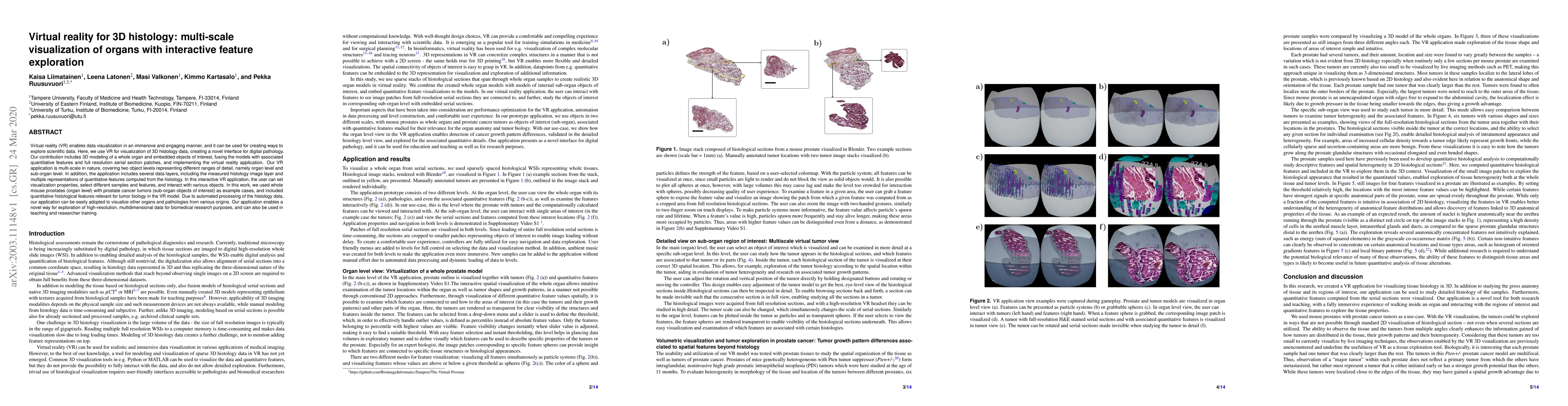
Virtual reality (VR) enables data visualization in an immersive and engaging manner, and it can be used for creating ways to explore scientific data. Here, we use VR for visualization of 3D histology data, creating a novel interface for digital pathology. Our contribution includes 3D modeling of a whole organ and embedded objects of interest, fusing the models with associated quantitative features and full resolution serial section patches, and implementing the virtual reality application. Our VR application is multi-scale in nature, covering two object levels representing different ranges of detail, namely organ level and sub-organ level. In addition, the application includes several data layers, including the measured histology image layer and multiple representations of quantitative features computed from the histology. In this interactive VR application, the user can set visualization properties, select different samples and features, and interact with various objects. In this work, we used whole mouse prostates (organ level) with prostate cancer tumors (sub-organ objects of interest) as example cases, and included quantitative histological features relevant for tumor biology in the VR model. Due to automated processing of the histology data, our application can be easily adopted to visualize other organs and pathologies from various origins. Our application enables a novel way for exploration of high-resolution, multidimensional data for biomedical research purposes, and can also be used in teaching and researcher training.
Get AI-generated insights about this paper's methodology, results, and significance.
Current paper (gray), citations (green), references (blue)
Enes Yigitbas, Dennis Wüppelman
Usman Alim, Roberta Mota, Ehud Sharlin
Sergei V. Bulanov, Marcel Lamač, Petr Valenta et al.
Karthik Ramani, Mayank Patel, Ananya Ipsita et al.
Daniela Ushizima, Ronald Pandolfi, Zineb Sordo et al.
Title | Authors | Year | Actions |
---|---|---|---|
The 3D Visualization of Digitalized Pathological Serial Sections on a Native Resolution
|
Miklós Vincze, Sándor Burian, Miklós Kozlovszky
|
2025
|
View Paper |
Monitoring the Examination of Digitised Pathology Samples in a 3D VR Environment
|
Miklós Vincze, Sándor Burian, Miklós Kozlovszky
|
2024
|
View Paper |
The potential of x‐ray virtual histology in the diagnosis of skin tumors
|
Giulia Saccomano, Maurizio Pinamonti, Elena Longo, Thomas Marcuzzo, Giuliana Tromba, Diego Dreossi, Francesco Brun
|
2024
|
View Paper |
Practical tips for teaching medicine in the metaverse
|
M. Rodriguez-Florido, Manuel Maynar
|
2024
|
View Paper |
Technologies for design-build-test-learn automation and computational modelling across the synthetic biology workflow: a review
|
R. Matzko, Savas Konur
|
2024
|
View Paper |
An end-to-end workflow for nondestructive 3D pathology.
|
K. Bishop, Lindsey A. Erion Barner, Qinghua Han, Elena Baraznenok, Lydia Lan, Chetan Poudel, Gan Gao, R. Serafin, Sarah S. L. Chow, A. Glaser, Andrew Janowczyk, David Brenes, Hongyi Huang, Dominie Miyasato, Lawrence D. True, Soyoung Kang, J. Vaughan, Jonathan T. C. Liu
|
2024
|
View Paper |
Engineering the future of 3D pathology
|
J. Liu, Sarah S. L. Chow, Richard Colling, M. Downes, Xavier Farré, Peter Humphrey, Andrew Janowczyk, T. Mirtti, Clare Verrill, Inti Zlobec, Lawrence D. True
|
2023
|
View Paper |
3D Visualization in Digital Medicine Using XR Technology
|
Miklós Vincze, B. Molnár, M. Kozlovszky
|
2023
|
View Paper |
An end-to-end workflow for non-destructive 3D pathology
|
K. Bishop, L. Barner, Qinghua Han, Elena Baraznenok, Lydia Lan, Chetan Poudel, Gan Gao, R. Serafin, Sarah S. L. Chow, A. Glaser, A. Janowczyk, David Brenes, Hongyi Huang, Dominie Miyasato, L. True, Soyoung Kang, J. Vaughan, Jonathan T. C. Liu
|
2023
|
View Paper |
Virtual Models Using Augmented Reality May Provide a Suitable Supplement, Although Not a Physical Specimen Replacement, in Pathology Education
|
Christian Moro, Dianheng Bu, Aditya Gadgil, Gordon Wright, Cindy J. Jones
|
2023
|
View Paper |
The HRA Organ Gallery affords immersive superpowers for building and exploring the Human Reference Atlas with virtual reality
|
Andreas Bueckle, Catherine Qing, Shefali Luley, Yash Kumar, Naval Pandey, Katy Börner
|
2023
|
View Paper |
Students’ Daily English Situational Teaching Based on Virtual Reality Technology
|
Lijuan Han
|
2022
|
View Paper |
A Brave New World: Virtual Reality and Augmented Reality in Systems Biology
|
Berk Turhan, Z. Gümüş
|
2022
|
View Paper |
Visualization in virtual reality: a systematic review
|
Elif Hilal Korkut, Elif Surer
|
2022
|
View Paper |
Spatial analysis of histology in 3D: quantification and visualization of organ and tumor level tissue environment
|
P. Ruusuvuori, Masi Valkonen, K. Kartasalo, Mira Valkonen, T. Visakorpi, M. Nykter, Leena Latonen
|
2022
|
View Paper |
Inspection of histological 3D reconstructions in virtual reality
|
Oleg Lobachev, Moritz Berthold, B. Steiniger, Henriette Pfeffer, M. Guthe
|
2020
|
View Paper |
Comments (0)