Virtual staining for 3D X-ray histology of bone implants

PDF

Publication

Published: Sep 11, 2025
Categories: eess.IV, cs.AI, cs.CV

Metrics

Source: ArXiv

Quick Actions

View PDF

Quick Answers

What methodology did the authors use?

The study employs a multi-modal image registration approach combined with a paired CycleGAN to generate 3D virtual histology from micro-CT and histological images. The model was trained on a dataset of co-registered micro-CT and histology whole slide images (WSIs) with various implant materials. More in Methodology →

What are the key results?

The model successfully generates high-resolution 3D virtual histology with accurate tissue segmentation and staining. — The approach enables the analysis of bone microstructure and implant degradation in 3D. More in Key Results →

Why is this work significant?

This research advances the field of digital pathology by enabling non-invasive 3D analysis of tissue samples, which has implications for better understanding implant integration and bone remodeling processes. More in Significance →

What are the main limitations?

The method requires co-registered micro-CT and histology data which can be challenging to obtain. — The model's performance is dependent on the quality and diversity of the training dataset. More in Limitations →

Paper Preview

Abstract

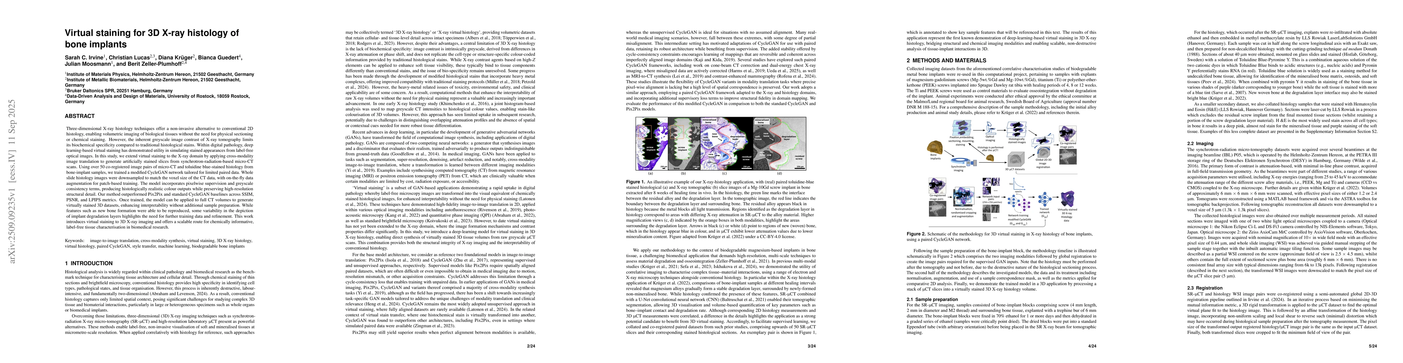
Three-dimensional X-ray histology techniques offer a non-invasive alternative to conventional 2D histology, enabling volumetric imaging of biological tissues without the need for physical sectioning or chemical staining. However, the inherent greyscale image contrast of X-ray tomography limits its biochemical specificity compared to traditional histological stains. Within digital pathology, deep learning-based virtual staining has demonstrated utility in simulating stained appearances from label-free optical images. In this study, we extend virtual staining to the X-ray domain by applying cross-modality image translation to generate artificially stained slices from synchrotron-radiation-based micro-CT scans. Using over 50 co-registered image pairs of micro-CT and toluidine blue-stained histology from bone-implant samples, we trained a modified CycleGAN network tailored for limited paired data. Whole slide histology images were downsampled to match the voxel size of the CT data, with on-the-fly data augmentation for patch-based training. The model incorporates pixelwise supervision and greyscale consistency terms, producing histologically realistic colour outputs while preserving high-resolution structural detail. Our method outperformed Pix2Pix and standard CycleGAN baselines across SSIM, PSNR, and LPIPS metrics. Once trained, the model can be applied to full CT volumes to generate virtually stained 3D datasets, enhancing interpretability without additional sample preparation. While features such as new bone formation were able to be reproduced, some variability in the depiction of implant degradation layers highlights the need for further training data and refinement. This work introduces virtual staining to 3D X-ray imaging and offers a scalable route for chemically informative, label-free tissue characterisation in biomedical research.

AI Key Findings

Generated Nov 01, 2025

Methodology — What approach did the authors take?

The study employs a multi-modal image registration approach combined with a paired CycleGAN to generate 3D virtual histology from micro-CT and histological images. The model was trained on a dataset of co-registered micro-CT and histology whole slide images (WSIs) with various implant materials.

Key Results — What are the main findings?

  • The model successfully generates high-resolution 3D virtual histology with accurate tissue segmentation and staining.
  • The approach enables the analysis of bone microstructure and implant degradation in 3D.
  • The method outperforms existing techniques in preserving histological details and spatial relationships.

Significance — Why does this research matter?

This research advances the field of digital pathology by enabling non-invasive 3D analysis of tissue samples, which has implications for better understanding implant integration and bone remodeling processes.

Technical Contribution — What is the technical contribution?

Development of a paired CycleGAN framework specifically tailored for generating 3D virtual histology from multi-modal biomedical imaging data.

Novelty — What is new about this work?

The work introduces a novel approach that combines advanced image registration with CycleGAN for 3D virtual histology generation, enabling detailed analysis of complex biological structures in three dimensions.

Limitations — What are the limitations of this study?

  • The method requires co-registered micro-CT and histology data which can be challenging to obtain.
  • The model's performance is dependent on the quality and diversity of the training dataset.

Future Work — What did the authors propose for future work?

  • Exploring hybrid architectures that incorporate both paired and unpaired data.
  • Improving color normalization techniques for more accurate virtual staining.
  • Extending the method to other tissue types and pathological conditions.

Paper Details

Paper ID: 2509.09235
Categories:
eess.IV cs.AI cs.CV physics.comp-ph q-bio.QM

How to Cite This Paper

BibTeX
@article{zellerplumhoff2025virtual,
  title         = {Virtual staining for 3D X-ray histology of bone implants},
  author        = {Zeller-Plumhoff, Berit and Guedert, Bianca and Lucas, Christian and others},
  year          = {2025},
  eprint        = {2509.09235},
  archivePrefix = {arXiv},
  primaryClass  = {eess.IV},
}
APA 7
Zeller-Plumhoff, B., Guedert, B., Lucas, C., Krüger, D., Moosmann, J., & Irvine, S. (2025). Virtual staining for 3D X-ray histology of bone implants. arXiv. https://arxiv.org/abs/2509.09235
MLA 9
Zeller-Plumhoff, Berit, et al. "Virtual staining for 3D X-ray histology of bone implants." arXiv, 2025, arxiv.org/abs/2509.09235.

Comments (0)

This website uses cookies to ensure you get the best experience. Learn more