Visibility of capillaries in turbid tissues: an analytical approach
Publication
Metrics
AI Quick Summary
This study develops an analytical model for visualizing capillary loops in turbid tissues using a perturbation approach for light propagation, providing explicit expressions for subsurface defect contrasts. The approach aids in designing and optimizing imaging systems for clinical applications like rheumatology and cancer detection.
Paper Preview
Abstract
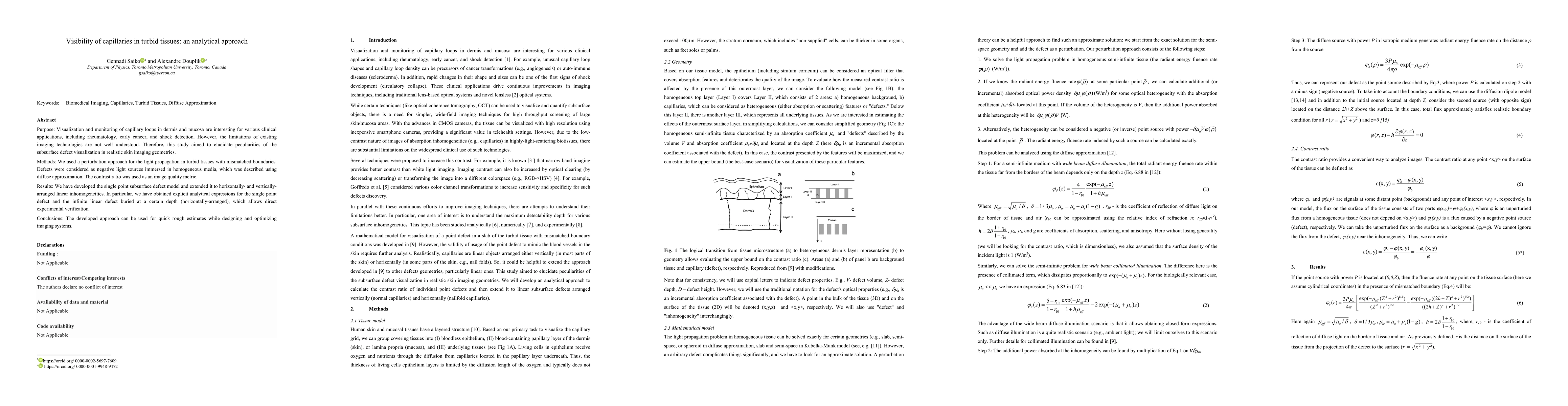
Purpose: Visualization and monitoring of capillary loops in dermis and mucosa are interesting for various clinical applications, including rheumatology, early cancer, and shock detection. However, the limitations of existing imaging technologies are not well understood. Therefore, this study aimed to elucidate peculiarities of the subsurface defect visualization in realistic skin imaging geometries. Methods: We used a perturbation approach for the light propagation in turbid tissues with mismatched boundaries. Defects were considered as negative light sources immersed in homogeneous media, which was described using diffuse approximation. The contrast ratio was used as an image quality metric. Results: We have developed the single point subsurface defect model and extended it to horizontally- and vertically-arranged linear inhomogeneities. In particular, we have obtained explicit analytical expressions for the single point defect and the infinite linear defect buried at a certain depth (horizontally-arranged), which allows direct experimental verification. Conclusions: The developed approach can be used for quick rough estimates while designing and optimizing imaging systems.
AI Key Findings
Get AI-generated insights about this paper's methodology, results, significance, and more — seven facets brought into focus.
Impact
Paper Details
Authors
PDF Preview
Key Terms
Citation Network
Current paper (gray), citations (green), references (blue)
Display is limited for performance on very large graphs.
Discussion 0