This paper introduces PATH-X, a framework combining Vision Transformers, Autoencoders, and SHAP for explainable AI to stratify cancer patient risk using Whole Slide Imaging (WSI). The method extracts and compresses features from WSIs, enabling effective clustering and classification, with SHAP providing interpretability by mapping key features onto histopathology images.
Cancer remains one of the leading causes of mortality worldwide, necessitating accurate diagnosis and prognosis. Whole Slide Imaging (WSI) has become an integral part of clinical workflows with advancements in digital pathology. While various studies have utilized WSIs, their extracted features may not fully capture the most relevant pathological information, and their lack of interpretability limits clinical adoption. 
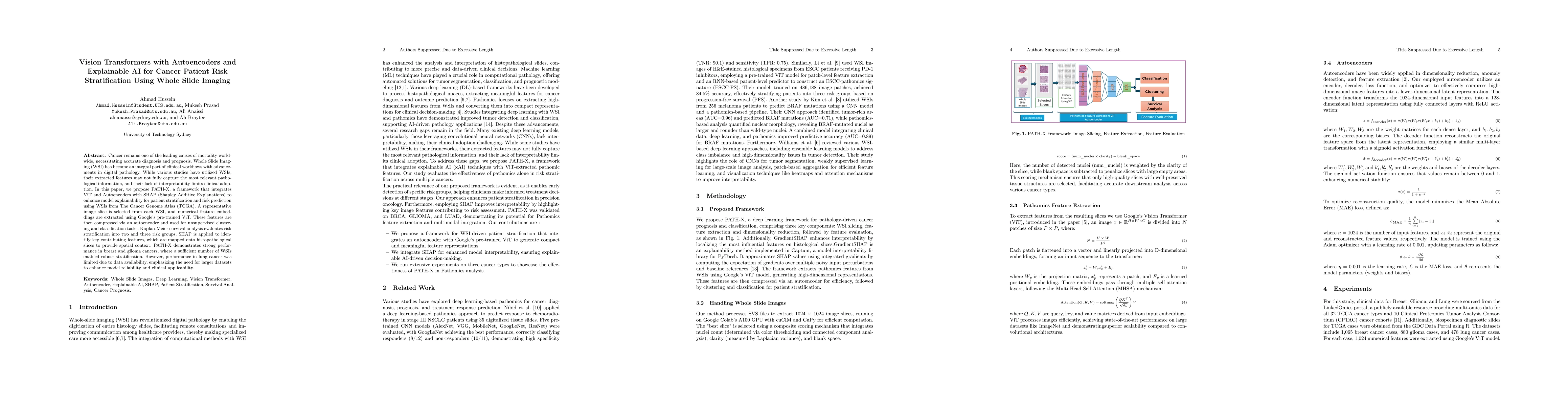
In this paper, we propose PATH-X, a framework that integrates Vision Transformers (ViT) and Autoencoders with SHAP (Shapley Additive Explanations) to enhance model explainability for patient stratification and risk prediction using WSIs from The Cancer Genome Atlas (TCGA). A representative image slice is selected from each WSI, and numerical feature embeddings are extracted using Google's pre-trained ViT. These features are then compressed via an autoencoder and used for unsupervised clustering and classification tasks. Kaplan-Meier survival analysis is applied to evaluate stratification into two and three risk groups. SHAP is used to identify key contributing features, which are mapped onto histopathological slices to provide spatial context. PATH-X demonstrates strong performance in breast and glioma cancers, where a sufficient number of WSIs enabled robust stratification. However, performance in lung cancer was limited due to data availability, emphasizing the need for larger datasets to enhance model reliability and clinical applicability.
Generated Jun 10, 2025
The research proposes PATH-X, a framework integrating Vision Transformers (ViT), Autoencoders, and SHAP for cancer patient risk stratification using Whole Slide Imaging (WSI) from TCGA. It involves selecting representative image slices, extracting numerical features using pre-trained ViT, compressing these features via autoencoders, and applying clustering and classification tasks. Kaplan-Meier survival analysis evaluates stratification, with SHAP identifying key contributing features mapped onto histopathological slices.
This research is significant as it enhances model explainability for cancer patient stratification using WSIs, aiding in accurate diagnosis and prognosis, which are crucial for effective cancer management.
The main technical contribution is the PATH-X framework that combines ViT for feature extraction, autoencoders for dimensionality reduction, and SHAP for model interpretability in cancer patient risk stratification using WSIs.
This work is novel as it integrates ViT, autoencoders, and SHAP for explainable pathomics analysis in cancer patient risk stratification, offering a more interpretable model compared to existing methods.
No key terms available for this paper.
Current paper (gray), citations (green), references (blue)
Jeroen van der Laak, Clément Grisi, Geert Litjens
Saeed Hassanpour, Manu Goyal, Jonathan D. Marotti et al.
Yang Chen, Jian Zhang, Yongbing Zhang et al.
Jin Tae Kwak, Anh Tien Nguyen, Keunho Byeon et al.
No citations found for this paper.
Comments (0)