This study demonstrates that X-ray Velocimetry can effectively visualize and quantify changes in lung ventilation following the placement of endobronchial valves in sheep, indicating potential for clinical use in emphysema treatment.
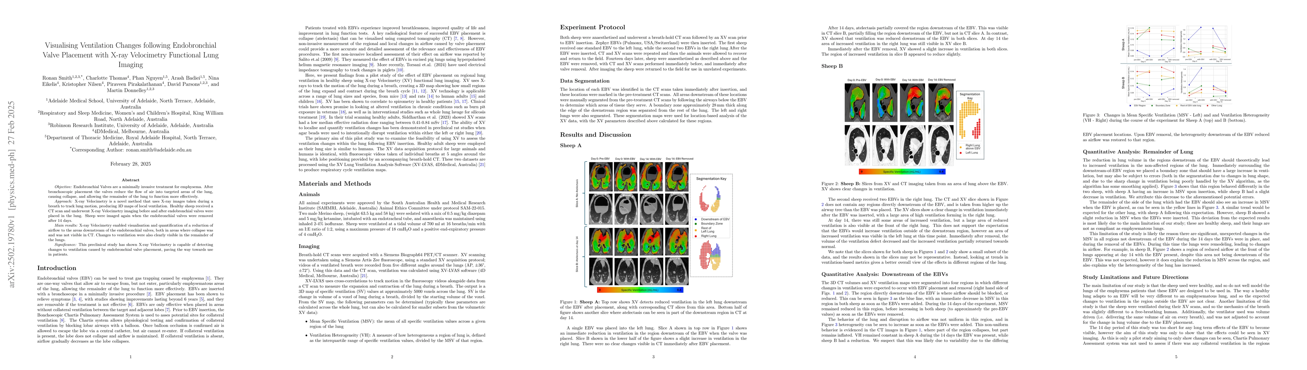
Objective: Endobronchial Valves are a minimally invasive treatment for emphysema. After bronchoscopic placement the valves reduce the flow of air into targeted areas of the lung, causing collapse, and allowing the remainder of the lung to function more effectively. Approach: X-ray Velocimetry is a novel method that uses X-ray images taken during a breath to track lung motion, producing 3D maps of local ventilation. Healthy sheep received a CT scan and underwent X-ray Velocimetry imaging before and after endobronchial valves were placed in the lung. Sheep were imaged again when the endobronchial valves were removed after 14 days. Main results: X-ray Velocimetry enabled visualisation and quantification of a reduction of airflow to the areas downstream of the endobronchial valves, both in areas where collapse was and was not visible in CT. Changes to ventilation were also clearly visible in the remainder of the lungs. Significance: This preclinical study has shown X-ray Velocimetry is capable of detecting changes to ventilation caused by endobronchial valve placement, paving the way towards use in patients.
Generated Jun 10, 2025
This study employed X-ray Velocimetry (XV) functional lung imaging, a novel method using X-ray images during breath holds to track lung motion, producing 3D maps of local ventilation. Healthy sheep underwent CT scans and XV imaging before and after endobronchial valve (EBV) placement, with follow-up scans after valve removal.
The study demonstrates XV's capability to detect ventilation changes due to EBV placement, paving the way for its use in emphysema patients.
The study presents X-ray Velocimetry as a promising technique for visualizing and quantifying ventilation changes in lungs after EBV placement.
This research is novel in its application of X-ray Velocimetry to study ventilation changes following endobronchial valve placement, offering a non-invasive, safe imaging technique for emphysema patients.
Current paper (gray), citations (green), references (blue)
Display is limited for performance on very large graphs.
M.-L., Smith, R., Reyne, N. et al.
Mehaffey, J Hunter, Hayanga, J W Awori, Luo, Xun et al.
David M. Paganin, Ying Ying How, Kaye S. Morgan et al.
No citations found for this paper.
Comments (0)