The study developed an AI using a 3D convolutional model of nnU-Net for automatic region segmentation of 4D-CT images during swallowing. The AI was trained with leave-one-out cross-validation and evaluated using Dice coefficients.
Visualization of Organ Movements Using Automatic Region Segmentation of Swallowing CT
Publication
Metrics
AI Quick Summary
This study introduces an AI model for automatic segmentation of 4D-CT images during swallowing, achieving high accuracy for most regions except for fast-moving structures. Future improvements are needed to enhance the precision and reliability of the AI’s segmentation capabilities.
Quick Answers
What is "Visualization of Organ Movements Using Automatic Region Segmentation of Swallowing CT" about?
This study introduces an AI model for automatic segmentation of 4D-CT images during swallowing, achieving high accuracy for most regions except for fast-moving structures. Future improvements are needed to enhance the precision and reliability of the AI’s segmentation capabilities.
What methodology did the authors use?
The study developed an AI using a 3D convolutional model of nnU-Net for automatic region segmentation of 4D-CT images during swallowing. The AI was trained with leave-one-out cross-validation and evaluated using Dice coefficients. More in Methodology →
What are the key results?
The AI achieved a median Dice coefficient of 0.7 or higher for the bolus, bones, tongue, and soft palate. — Regions like the thyroid cartilage and epiglottis had Dice coefficients below 0.7 due to rapid movement and metal artifacts. More in Key Results →
Why is this work significant?
This research significantly reduces labor and time required for segmentation, enabling processing of numerous cases in the future, and contributes to advancing the analysis of organ movements during swallowing and improving swallowing CT accuracy. More in Significance →
What are the main limitations?
Challenges include improving AI accuracy, especially for structures with low clarity in swallowing CT such as the hyoid bone, thyroid cartilage, and epiglottis. — Metal artifacts and motion blur in CT imaging pose ongoing challenges in improving image quality. More in Limitations →
Abstract
This study presents the first report on the development of an artificial intelligence (AI) for automatic region segmentation of four-dimensional computer tomography (4D-CT) images during swallowing. The material consists of 4D-CT images taken during swallowing. Additionally, data for verifying the practicality of the AI were obtained from 4D-CT images during mastication and swallowing. The ground truth data for the region segmentation for the AI were created from five 4D-CT datasets of swallowing. A 3D convolutional model of nnU-Net was used for the AI. The learning and evaluation method for the AI was leave-one-out cross-validation. The number of epochs for training the nnU-Net was 100. The Dice coefficient was used as a metric to assess the AI's region 
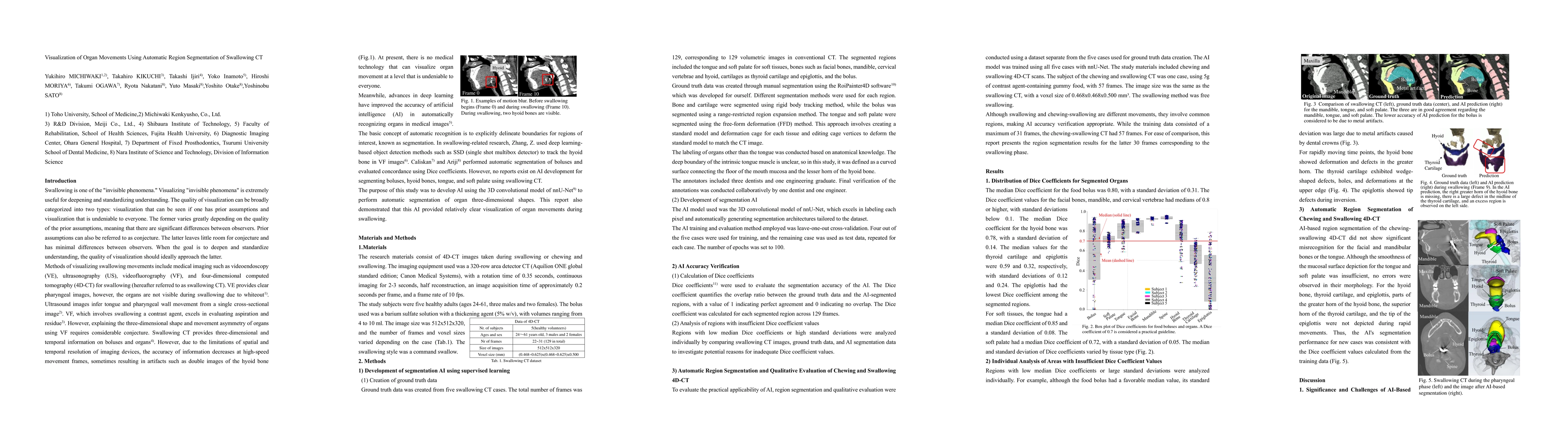
segmentation accuracy. Regions with a median Dice coefficient of 0.7 or higher included the bolus, bones, tongue, and soft palate. Regions with a Dice coefficient below 0.7 included the thyroid cartilage and epiglottis. Factors that reduced the Dice coefficient included metal artifacts caused by dental crowns in the bolus and the speed of movement for the thyroid cartilage and epiglottis. In practical verification of the AI, no significant misrecognition was observed for facial bones, jaw bones, or the tongue. However, regions such as the hyoid bone, thyroid cartilage, and epiglottis were not fully delineated during fast movement. It is expected that future research will improve the accuracy of the AI's region segmentation, though the risk of misrecognition will always exist. Therefore, the development of tools for efficiently correcting the AI's segmentation results is necessary. AI-based visualization is expected to contribute not only to the deepening of motion analysis of organs during swallowing but also to improving the accuracy of swallowing CT by clearly showing the current state of its precision.
Key Findings, in focus
Seven facets of this paper, analysed and brought into focus by AI.
This research significantly reduces labor and time required for segmentation, enabling processing of numerous cases in the future, and contributes to advancing the analysis of organ movements during swallowing and improving swallowing CT accuracy.
- The AI achieved a median Dice coefficient of 0.7 or higher for the bolus, bones, tongue, and soft palate.
- Regions like the thyroid cartilage and epiglottis had Dice coefficients below 0.7 due to rapid movement and metal artifacts.
- Practical verification showed no significant misrecognition for facial bones, jaw bones, or the tongue, but hyoid bone, thyroid cartilage, and epiglottis were not fully delineated during fast movements.
This research significantly reduces labor and time required for segmentation, enabling processing of numerous cases in the future, and contributes to advancing the analysis of organ movements during swallowing and improving swallowing CT accuracy.
The development and application of nnU-Net for automatic region segmentation of 4D-CT images during swallowing, showcasing its effectiveness in reducing manual segmentation time significantly.
This work is novel in presenting the first AI for automatic region segmentation of swallowing CT, providing a practical solution for reducing labor and time in manual segmentation while highlighting areas for future improvement in AI accuracy and correction tools.
- Challenges include improving AI accuracy, especially for structures with low clarity in swallowing CT such as the hyoid bone, thyroid cartilage, and epiglottis.
- Metal artifacts and motion blur in CT imaging pose ongoing challenges in improving image quality.
- Increasing ground truth data and exploring evaluation methods beyond Dice coefficients to improve AI's region segmentation accuracy for tongue and food bolus.
- Developing tools for efficiently correcting AI-generated segmentation results to mitigate the risk of misrecognition.
Paper Details
Authors
PDF Preview
Related Papers
No references found for this paper.
Discussion 0